
Journal of Veterinary Science & Medicine
Download PDF
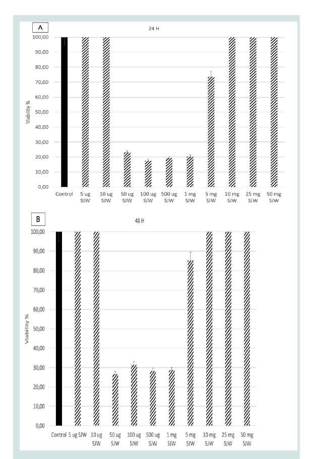 Figure 1: Cell viability results for SJW at a) 24h b) 48h c) 72 h incubation
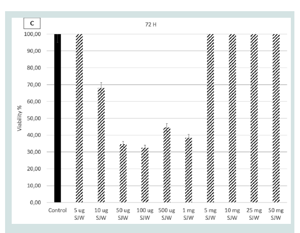 Figure 2: Tumor sizes of the 5 experimental groups. a) Control Group b)
Doxorubicin Group. c) Group to examine the slowing effect of SJW (when
tumor size reaches 0.2 cm, SJW will be applied) d) To study the treatment
effect of Group SJW (when tumor size reaches 0.8 cm, SJW will be applied)
e) Doxorubicin (5mg/kg) and SJW combination] before sacrification.
Figure 2: Tumor sizes of the 5 experimental groups. a) Control Group b)
Doxorubicin Group. c) Group to examine the slowing effect of SJW (when
tumor size reaches 0.2 cm, SJW will be applied) d) To study the treatment
effect of Group SJW (when tumor size reaches 0.8 cm, SJW will be applied)
e) Doxorubicin (5mg/kg) and SJW combination] before sacrification.
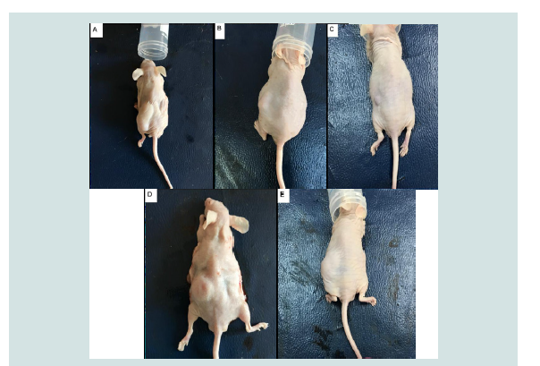 Figure 3: Histopathology of tumors: a) Control group: no necrosis; b) DOX
group: prominent necrosis; c) SJW early: prominent necrosis cell death
appears; d) SJW late: prominent necrosis cell death appears; e) DOX+SJW:
prominent necrosis cell death appears.
Figure 3: Histopathology of tumors: a) Control group: no necrosis; b) DOX
group: prominent necrosis; c) SJW early: prominent necrosis cell death
appears; d) SJW late: prominent necrosis cell death appears; e) DOX+SJW:
prominent necrosis cell death appears.
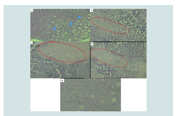 Figure 4: a). Viable tumour tissue in control mouse. The wide arrow
represents invasive border. b). The red ellipse represents prominent necrosis
area. c). The red ellipse represents prominent necrosis cell death area. d).
The red ellipse represents prominent necrosis cell death area. e). All of the
are represents necrotic tissue.
Figure 4: a). Viable tumour tissue in control mouse. The wide arrow
represents invasive border. b). The red ellipse represents prominent necrosis
area. c). The red ellipse represents prominent necrosis cell death area. d).
The red ellipse represents prominent necrosis cell death area. e). All of the
are represents necrotic tissue.
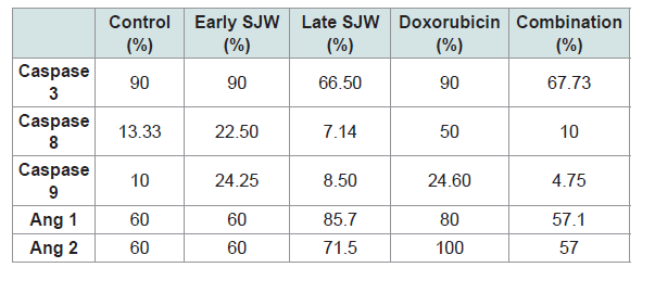 Table 1: İmmunohistochemical Parameters
Research Article
Investigation of In-Vivo Effect of St. John’s Wort (Hypericum Perforatum) In Lung Cancer
Aktaş S*, Erol A, Serinan EO, Gökbayrak OE and Altun Z
Department of Basic Oncology, Institute of Oncology, Dokuz Eylul University, Izmir, Turkey
*Address for correspondence:
Aktaş S, Department of Basic Oncology, Institute of Oncology, Dokuz
Eylul University, Izmir, Turkey; Email: safiyeaktas@gmail.com
Submission: 7 July, 2021;
Accepted: 10 August, 2021;
Published: 15 August, 2021
Copyright: © 2021 Aktaş S et al. This is an open access article
distributed under the Creative Commons Attribution License, which
permits unrestricted use, distribution, and reproduction in any medium,
provided the original work is properly cited.
Abstract
Due to limitations in the treatment of lung cancer, finding natural
compounds from plants can provide an alternative treatment for lung
cancer. St. John’s Wort (SJW) has anti-proliferative and pro-apoptotic
properties that can be used in lung cancer treatment. The aim of
this study is to explore antitumor effect of SJW in lung cancer in vivo
animal model. 35 animals; 7 animals in each group were randomized
as control, Doxorubicin, SJW early treatment, SJW treatment, and
doxorubicin+SJW groups. After 7 days sacrification was performed.
Tumor diameter did not show statistically significant change but in
all four-group compared with control group; tumor tissue showed
prominent necrosis and apoptosis. No histologic changes observed in
other tissues. Biochemistry did not show organ insufficiency.
SJW is shown to have antitumoral effect in subcutaneous xenograft
lung cancer in vivo model in nude mice. Dose was obtained
comparing with DOX. In combination with DOX, there were no
synergistic increase in anti-tumo effect. SJW might be a candidate
antineoplastic supplementation in lung cancer.
Introduction
Lung cancer is one of the most common malignant tumors in the
world, consisting of pathologically and clinically diverse subtypes [1-4]. Due to limitations in the treatment of lung cancer, finding natural
compounds from plants can provide an alternative treatment for lung
cancer [5,6]. St. John’s Wort (St. John’s Wort = SJW = Hypericum
Perforatum) is also one of these plants used. SJW has previously taken
its place in the literature as a herbaceous herb used in the treatment of
fibrosis, neuralgia, depression and anxiety as an alternative to classical
antidepressants [7]. SJW’s most well-known bioactive compounds
are hypericin, hyperoside and hyperforin. Hypericin; SJW started to
take part in cancer studies after it was revealed that reactive oxygen
species (ROS) are produced in cells and photodynamic therapy (PDT)
that induces apoptosis, necrosis or autophagy is a photo-sensitizer
that can be used [7,8]. After the use of SJW in cancer research, it has
also been revealed that the accumulation of hypericin in neoplastic
tissue is significantly higher than normal tissue and can be used as
an effective fluorescent marker for tumor detection and imaging
in photodynamic diagnosis (PDD) [8]. However, drug interaction
of hypericin and hyperforin, which are active components of SJW,
with anticancer drugs have been reported in various studies. SJW
modulates the expression of multidrug resistance-1 (MDR-1), which
is the main multidrug resistance mechanism responsible for the
failure of chemotherapy [8].
SJW has anti-proliferative and pro-apoptotic properties that can
be used in lung cancer treatment [7,8]. The proliferation inhibitory
effect and apoptosis-inducing effect of hyperoside in lung cancer have
been shown in various studies [9,10]. Although there are studies in
the literature showing the anti-proliferative and pro-apoptotic effects
of SJW in many different types of cancer, there is no study evaluating the combination of SJW with an agent used in traditional lung cancer
treatment.
In the in-vitro part of the study, the 5μg/ml-50mg/ml dose range
of SJW on the LLC lung cancer cell line was applied at 24, 48, 72 hours
incubation times and WST-1 cell viability test was performed. The
LD50 value of SJW was determined to be 50 μg / ml for 24 hours.
The aim of this study is to investigate the anti-cancer effects of
SJW (hypericin) an in vivo experimental animal model of lung cancer.
Materials and Methods
This study was approved by Dokuz Eylul University Local Ethics
Commitee for Multidisciplinary animal research by number 28/2019.
Nature’s Bounty St. John’s Worth: Hypericum perforatum
(over earth), includes Hyipericin 0.9 mg (0.3%) in one capsule and
Doxorubicin (KOÇAK) (10 mg/5 ml) are the chemicals used.
Cell Culture based in-vitro studies:
Lewis Lung Carcinoma (LLC) (ATCC, CRl-1642) cell line was
cultured in RPMI 1640 +10 % FBS+ 1 %Penicillin/Streptomycin + 1
%L-glutamin at 5% CO2 37°C.
Doxorubicin (5mg/ kg) and SJW (5μg/ml-50mg/ml) doses
were applied to 96 well plate with 6 wells of each condition at 24,
48 ve 72 hours. Proliferation was assessed with WST-1 at 450/630 by
ELISA raeader [11]. Extracellular migration and invasion test were
performed by invasion chamber in 24 well plate with polycarbonate
membrane.
In Vivo Xenograft tests were performed using 35, 5-7 weeks old
male nude mice average 25 grams in 5 groups (7 mice in each group)
as follows:
1) Control group 0.9 cm tumor, (IP 0.3 cc saline)
2) Doxorubicin group (5mg/kg) (IP in 0.3 cc)
3) Group to examine the slowing effect of SJW (when tumor size
reaches 0.2 cm, 50 ug SJW was applied)
4) To study the treatment effect of Group SJW (when tumor size
reaches 0.9 cm, 50 ug SJW was applied)
5) Doxorubicin (5mg/kg) and 50 ug SJW combination
The animals were kept in HEPA filtered cabinets at standard
conditions (20 ± 2 ºC) room temperature and 12 hours day/night
cycle. They were fed by sterile water and pellet ad abitum. After
5 days 4x10⁶ LLC cells in 0.3 cc RPMI were injected to left flanck.
Daily observation, weight control, tumor diameter control was done
till sacrification. When tumors reached to 0.2 and 0.9 cm in greatest
diameter, they were randomized to groups.
After 7 days animals were sacrified under Halotan anestesia
(Halotan BP 250 ml Pirimal). Whole blood from vena cava inferior
and urine from the bladder were aspirated. Strip urine test was
applied. The blood near 0.5 cc each was immediately centrifugated
at 2000 x G in microtube. Supernatant serum was seperated and kept
at -20 C till biochemistry. Serum glucose (mg/dL), creatinine (mg/
dL), AST(U/L), ALT(U/L) was calculated by spectrophotometric
analysis with IVD veterinary Preventive Care Profile Plus kit
(ABAXIS, Germany) at Vetscan VS2 Chemistry Analyser (Abaxis)
at Dokuz Eylul University Izmir Health Technologies Development
and Accelerator Center (BioIzmir). Calibration was done by Abaxis
Control-I (Randox) kit. Urine tests microalbumin and creatinine
were done by IVD Clintek Microalbumin kit (SIEMENS, Germany)
with Clintek Status Analyzer (Siemens) at BioIzmir. Check-stix
Combo (SIEMENS, Germany) kit was used for calibration. Tumor
tissue, lung, kidneys, heart, brain, liver were kept in 10% neutral
formaline and then embedded in parafin. Tissue sections were stained
with hematoxylin & eosin and TUNEL, Caspase 3,8,9, Ang1, Ang 2
immunohistochemistry (IHC) were performed to tumor tissues.
Apoptosis rate was determined by IHC. To do this, tissues were
stained with Caspase-3, Caspase-8, and Caspase-9 proteins. Sections
from cassettes obtained from tissues were first deparaffinized and
treated with 3% H2O2. The washing step was carried out with distilled
water and PBS for 10 min. Then tissues were treated Blocker A for 5
minutes and Blocker B for 5 minutes. The primary antibodies used
were diluted 1:200 and the slides were treated with the antibody for
1 hour. After primary antibody, all slides were washed with PBS for
10 min. Secondary antibody was added and left for 30 min. After
washing with PBS, DAB dye and H2O2 were added and left for 30
minutes. Slides were washed with tap water and PBS, respectively.
Slides were treated with Copper D and waited for 4 minutes. Then,
all slides were washed with tap water and stained with hematoxylin
and eosin dye. Slides, which were washed again with tap water, were
treated with a bluing reagent for 5 minutes. Then all slides were
washed with tap water again. Finally, all slides were subjected to a
series of increasing alcohol and treated with xylol for 1 hour. Before
the xylol was completely dry, the slides were covered with a coverslip
with Entellan and subjected to microscopy.
Results
WST-1 cell proliferation tests showed that LD50 for SJW is 50 μg/
ml (Figure 1). When the experimental results were evaluated, it was
observed that invasion and migration decreased significantly in 24
hours in the SJW group and 48 hours in the combined drug group.
When SJW was applied to a 9mm tumor, no reduction in tumor size
was observed (Figure 2). However, it caused necrotic cell death in
tumor tissue (Figure 3). When comparing the control group with SJW,
it was observed that tumor cell necrosis was statistically different in the
SJW group. This necrotic effect was observed both on 9 mm tumors
and when SJW was given after 0.5c2 mm tumor formation. Similar
tumor necrosis was observed in the SJW + DOX group. However, no
synergistic effect was observed in the combination of SJW with DOX.
Although the apoptotic effect is higher in the combination group than
the control group, the necrotic effect is lower than the control group.
Tunel Assay Results:
The mean apoptosis ratio for all groups was 37.38% ± 5.701 (0-
20). The mean apoptosis ratio was 3.20% ± 2.683 (0-6) in the control
group, 6,00 % +- 4,183 (0-10) in the Doxorubicin group, 15.00 % +-
5.774 (10-20) in the late SJW group, 8.20% +- 6.834 (3-20) in the early
SJW group and 6.00% +- 2.236 (5-10) in the combination group.
The highest apoptosis rate was observed in Late SJW group.
Combination did not caused synergistic effect for apoptosis.
This apoptotic effect occurred both through intrinsic and extrinsic pathway. İmmunohistochemical parameters are given in Table 1. SJW application increased Ang1 expression slightly, while
DOX application increased Ang1 expression more. Ang2 expression
has not changed. In biochemical blood results only, AST levels
increased in SJW + DOX group, while other test results are similar
among the groups. As a result, SJW has been shown to have an antitumoral
effect similar to DOX in the in-vivo experimental animal
subcutaneous lung cancer model. No synergistic effect was observed
in application with DOX
Discussion
The anticancer effect of SJW on a wide variety of cancer types
has been studied. Liu et al. showed that hyperoside, one of the active
ingredient of SJW, exerted inhibitory role in lung cancer development
[9]. Yang et al. showed that hyperoside significantly inhibited the
viability of lung cancer cells in a time- and dose-dependent manner
and enhanced the percentage of apoptotic cells [10]. In our study,
hypericin, another active ingredient of SJW, was studied. Hypericin
photodynamic therapy (PDT) efficacy has been studied in a mouse
tumor model. In the study, the primary mechanism of hypericinmediated
PDT mechanism stems from vasculature damage [8].
The mechanism of apoptotic process due to photodynamic
therapy of hypericin in Jurkat cells has also been studied. The
treatment also increases the activity of caspase-8 and caspase-3 and
increases apoptosis, which can be blocked by caspase-8 (Z-IETDFMK)
and caspase-3 (Z-DEVDFMK) inhibitors [8]. SJW ethanol
extract inhibited cell growth in a dose-dependent manner, as in
ethanol extract, in an apoptosis-induced manner [7]. In addition, this
SJW extract inhibits the AMPK / mTOR pathway, causing an increase
in the expression of pro-apoptotic proteins BAX and BAD, and a
decrease in the expression of anti-apoptotic proteins BCL-2, BCL-XL
[7]. In cell leukemia lines (K562 and U937) made by Hostanska et al.,
Hypericin and hyperforin, which are the active ingredients of SJW,
have synergistic effects, hyperforin induces apoptosis in doses, and
proliferation of hyperforin (K562 and U937) [12]. In a device made
by Stavropoulos et al., The effect of SJW, which has photostotoxic
effect in cancer, on cell proliferation was investigated in vitro. In the
study, when applied with 4-8 J / cm2 laser application on SJW cells
in 60 ug / ml sul, the execution head inhibited cell proliferation more
than 80% [13].
In the study of Borawska et al., It was found that SJW application
in the Glioblastoma cell line (U87MG) inhibits proliferation and
migration depending on the dose and time; and its anti-proliferative
and anti-migration properties have been shown to increase
synergistically with other herbal components such as propolis [14].
In the study conducted by Mirmalek et al., SJW, which has a cytotoxic
effect, was used in addition to Cisplatin in the MCF-7 breast cancer cell
line to overcome the chemotherapy resistance seen in breast cancer.
In the study, SJW has been shown to decrease cell proliferation in a
dose and time dependent manner. It has been shown that SJW, whose
LD50 dose is 5 μg / ml in 24 hours incubation, causes 60% apoptosis
in MCF-7 cells. The fact that it causes an increase in BAX expression
and a decrease in BCL-2 expression supports the apoptosis-inducing
effect of SJW [15]. In the study of Dona et al., the anticancer properties
of SJW in a wide variety of cell lines were investigated. Fibrosarcoma
(HT-1080), neuroblastoma (SK-N-BE), Melanoma (B16-LU8),
adenocarcinoma (c-26) cell lines were used in the study, and SJW was
shown to inhibit cell proliferation in a dose-dependent manner (IC50
value is 5). It varies from cell to cell between 8 umol / l). As a result
of the study, it was shown that hyperforin, the active ingredient of
SJW, can be an effective herbal agent in preventing cancer invasion
and metastasis [16].
The effects of SJW have also been examined in lung cancer, and
the majority of studies have been conducted in-vitro. In studies using
the non-small cell lung cancer cell line A549, it has been shown that
the active ingredients of St. John’s wort, hyperforin and hypericin,
induce autophagy and apoptosis, regulate caspase-3, caspas-9, and
p53-related cell death, depending on dose and time [17-19]. All these
results show that SJW extract has anti-proliferative and pro-apoptotic
effects.
Conclusion
In conclusion SJW is shown to have antitumoral effect in
subcutanous xenograft lung cancer in vivo model in nude mice. Dose
was obtained comparing with DOX. In combination with DOX, there
were no synertistic increase in anti-tumor effect. SJW might be a
candidate antineoplastic supplementation in lung cancer. Since this
is an acute study, the treatment response to the agent administered
within the first 7 days is interpreted by agent-induced cell death
within the tumor rather than reduction in tumor size.
Acknowledgement
This study was supported by Dokuz Eylul University Scientific
Research Committee by Project number: 2020.KB.SAG.002.
All animal experiments comply with the ARRIVE guidelines and
carried out in EU Directive 2010/63/EU for animal experiments
References
Citation
Aktaş S, Erol A, Serinan EO, Gökbayrak OE, Altun Z. Investigation of In-Vivo Effect of St. John’s Wort (Hypericum Perforatum) In Lung Cancer. J Veter Sci Med. 2021;9(1): 5