
Journal of Veterinary Science & Medicine
Download PDF
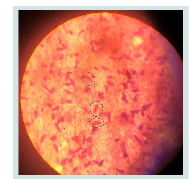 Figure 1: Sarcocystisic bradyzoites stained by Giemsa with 1000X
magnification.
Figure 1: Sarcocystisic bradyzoites stained by Giemsa with 1000X
magnification.
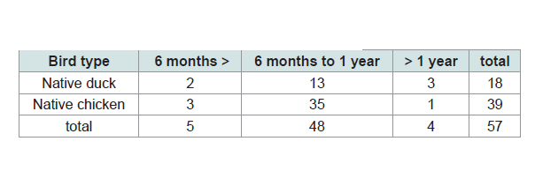 Table 1: Bird species categorized by different ages.
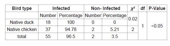 Table 2: Percentage and Number of different infected species of birds with
Sarcocystis.
Table 2: Percentage and Number of different infected species of birds with
Sarcocystis.
 Table 3: The relationship between the age of the birds and the prevalence of Sarcocystis.
Research Article
Investigation of Parasitic Sarcocystis Infection in Native Poultry Carcasses in North Part of Iran, Mazandaran (Amol)
Vahedi Noori N1*, Salehi A2, Razavi M2 and Masoumi M2
1Agricultural Research, Education and Extension Organization
(AREEO), Iran
2Veterinary Medicine Student, Babol Islamic Azad University, Iran
*Address for Correspondence: Vahedi Noori N, Assistant Professor, Razi Vaccine and Serum Research Institute, Agricultural Research, Education and Extension Organization (AREEO), Karaj, Iran; E-mail: nsvahedi@yahoo.com
Submission: 24-October, 2019
Accepted: 03-November, 2019
Published: 04-November, 2019
Copyright: © 2019 Vahedi Noori N, et al. This is an open access article
distributed under the Creative Commons Attribution License, which
permits unrestricted use, distribution, and reproduction in any medium,
provided the original work is properly cited.
Abstract
Sarcocystis is one of the most important and common protozoan
parasites in the world. Various species of Sarcocystis reported in groups of
mammals, birds and reptiles. In the life cycle of these parasite there are
2 hosts including hunted and hunter. Usually, omnivores and herbivores,
as intermediate hosts (hunted) and carnivores, are considered as the
definitive host (hunter) of this parasite. This research for the first time
examines the contamination of Sarcocystis (microcyst) in native birds of
Mazandaran province (Amol city). For this purpose, randomly, 57 native
bird’s breast muscles which include 18 pieces of native ducks and 39
native chickens were tested by digestion method. The results of the
experiment showed that 55 cases (96.5%) were infected with Sarcocystis
bradyzoite that contributed 100% to the local duck and 94.78% to the
native species. Based on age groups, the percentage of infection in the
group age under 6 months was 80%, in the age between 6 months and
one year was 97.91% and in the age group over one year, was 100%. The
Chi-square test did not show a significant difference in the percentage
of infection between two types of birds (duck-chicken) and age groups
(P <0.05).
Keywords
Contamination; Sarcocystis; Poultry carcasses; Amol
Introduction
The parasitic members of the genus Sarcocystis are coccidia
protozoa belonging to the Sarcocystidae family that cause
intracellular cysts. This family currently contains more than 220
species [1]. These parasites have 2 obligatory hosts in their life cycle,
including intermediate and definitive. Vegetarians and omnivores are
commonly referred to as intermediate hosts (hunted) and carnivores
as the definitive hosts (hunter) of this parasite. Asymptomatic
proliferation of the parasites is mediated by hosts, followed by
division of the merogenic cysts in the muscles. The parasite Sexual
stage, which is associated with the formation of oocysts or sporocysts,
occurs in the definitive host intestine [2], and reported in a variety
of Sarcocystis species in mammalian, avian and reptile groups.
Sarcocystis are able to carry out sexual and asexual reproduction in
a host [3]. DNA analysis and parasitological morphological studies
indicate that some of the species are present in at least two different
intermediate host [4,5]. Some of sarcocyte species are pathogenic
for humans and domestic animals and cause Sarcocystisosis. The
parasitic pathogen is mainly caused by intermediate hosts and is
mild in the definitive host. The rate of complications of this parasite
depends on factors such as the species, the severity of the infection
and the location of the parasite in the body. Pregnancy, lactation,
stress and lack of nutrients can increase the severity of the parasitic
pathogenesis [6,7]. So far, about 30 species of Sarcocystis have
identified in birds that produce cysts in at least thirteen orders of the
bird [4]. The definitive host of two species, Sarcocystis Wenzley and
Sarcocystis Horwath in chickens, are dogs and cats [8]. For other bird
species, the Sarcocystis species did not mentioned. In North America,
large Sarcocystis cysts have identified in goose and duck [9]. These macrocysts attributed to Riley’s Sarcocystis, which resemble rice grains
[10,11]. The wild duck has also been introduced as an intermediate
host for this protozoan. It seems in the protozoan life cycle, there
are more intermediate hosts [12]. Because of Sarcocystis’s mild
pathological complication, contaminated bird’s meat is unsuitable
for food consumption [13,14]. In wildlife, Sarcocystis contamination
occurs frequently. Sarcocystis falcachula, which is the ultimate host
of the eposome and the intermediate host of sparrows and native
poultry, can cause disease in domestic birds living in an open and
caged environment [15]. However, strains of Sarcocystis recognized
as infectious agents in domestic poultry around the world but they are
usually less pathologically important. Cysts caused by this protozoan
in intermediate hosts are large (macrocystic) or small (microcystic)
depending on the species and definitive host of the parasite. If the
cysts are large, they can easily diagnosed but if the cyst is small, the
diagnosis is impossible and the parasite easily enters the human food
cycle or other carnivorous organisms. In Iran, research on Sarcocystis
contamination in poultry, unlike ruminants, is infrequent. Similarly,
in a randomized study of pigeons infected with the nematode Hagyla
Trankata, the Sarcocystis was first isolated and identified from
the muscular layer of its gizzard [16]. This study for the first time
investigates the contamination of Sarcocystis (Microcyst) in native
birds of Mazandaran province (Amol city).
Materials and Methods
The method used in this study is observational and analyticalsectional.
For this purpose, 57 native bird species (native duck and
native chickens) were selected at random. Table 1 shows the number
and age of each bird studied. After slaughter, samples were taken
from each bird’s breast muscle for testing. Samples were analyzed
by the digestive method of Dobby et al. [17]. For this purpose, first
select 20 g of each sample and after grinding, with 100 ml of digestive
solution including: 10 ml of 32% sulfuric acid plus 2.5 g of pepsin
powder (Merck 7185 and 0.7 PIP-u/g) Mixed in one liter of distilled
water and place in a hot water bath at 37 °C for 30 minutes. After this
time and tissue digested, the samples were refined using a two-layer
filter. The obtained solutions were centrifuged at 1500 rpm for 10 min
and the precipitates were prepared on slides of monotonic spreads
and fixed with methanol after drying. At last, the slides were stained with 10 percent Giemsa and examined by light microscope. SAS 9/4
software and chi-square test with 95% confidence level (P <0.05) were
used to compare the frequency of infection in the studied bird species
and to compare the percentage of infection in different age groups.
Results
In this study, a total of 57 native bird species including 18 native
ducks and 39 native chickens were studied (Table 1). The results of
digestion experiments on the samples showed that 55 (96.5%) were
infected with Sarcocystis bradyzoite (Figure 1), and the percentage
of contamination in native ducks, was 100% and in native chickens,
94.78% (Table 2). The studied birds were categorized as under
6 months, 6 months to one year and over one year in (Table 1).
Accordingly, the infection rate in the age group under 6 months was
80%, in the age group of 6 months to one year, 97.91% and in the age
group above one year was 100% (Table 3).
Discussion and Conclusion
Sarcocystisosis is one of the most common protozoan parasitic diseases in the world. This study for the first time examines the
microcysts in native poultry muscles of Mazandaran province (Amol
city). For this purpose, 57 native poultry muscles including 18 native
ducks and 39 native chickens were tested. Although 11 rural birds
infected with Sarcocystis have been studied in three cases with acute
pulmonary symptoms, in five cases with musculoskeletal disease
and in three others with neurological symptoms [18], in our study
no clinical signs was recorded and didn’t observed in the studied
birds. Based on the results, 96.5% of all studied samples infected
with Sarcocystis (Table 2). The results of 191 chickens, 514 ducks
and 9 pigeons showed that only 17 (9.8%) of the studied chickens
had Sarcocystis isolated from their nervous system and identified
but in other species (ducks, pigeons) no parasites observed. Results
of poultry survey in central Nigeria showed that 3 out of 40 poultry
infected with Sarcocystis [19]. Surveys of native birds in New Zealand
have shown 11% of Sarcocystis infection [20]. Lithuania’s results
showed that only one of the 97 poultry (21 turkeys and 76 poultry)
was infected bySarcocystis [21]. Comparison of the results of this
study with the results of other researchers in different parts of the
world proves that the infection of this protozoan in native poultry
of Mazandaran province is at high rate. Since the identification of
parasite’s species and their definitive hosts were not considered in
this study, therefore, irrespective of the type of parasitic species and
their definitive hosts, the main reasons for this may be due to the
presence of suitable parasitic species and the diversity of the definitive
hosts. Our study area, together with other environmental factors,
has provided the appropriate conditions for this protozoan activity.
However, this requires substantial research in this area.
Based on the results all of the studied ducks (100%) were infected
with the Sarcocystis protozoa, which is higher than the percentage of
indigenous chicken (94.78%) (Table 2). Chi-square test showed no
significant difference between infection rates between the two groups
(native duck - native chickens) (P <0.05). However, the reason for this
difference may depends on the environment and the way the ducks
live. Basically, ducks live in humid and abundant water. This makes it
easy for the definitive host to excrete the stool and spread the parasite.
Therefore, the contamination is higher than other native chickens.
Research shows that ducks are more likely to be infected than other
birds due to direct and permanent contact with muddy and sludge
fields along with the excretion of definitive hosts or contaminated
meats containing adult cysts [9].
According to the results of this study, the percentage of infection
in different age groups in native ducks was 100% and there was no
difference between them (Table 3). Whereas in the studied poultry,
the percentage of infection was different in different age groups and
the percentage of contamination increased with increasing age of the
poultry (Table 3). Chi-square test showed no significant difference
between infection rates among different age groups (P <0.05). Also,
this difference was not significant in the studied poultry (native
duck - native poultry) (P <0.05). In one study of poultry, Sarcocystis
infection in under eight weeks’ sold group was zero and in over eight
Weeks’s group was 7.5% [19]. Although with age, the likelihood of
getting involved with infectious agents increases but due to the short
life span of the parasite [22], this difference is not significant in our
age groups with a range of six months.
Infectious Sarcocystisis an opportunistic infection that can be
easily manifested in people with AIDS or immunocompromised
patients [23]. We hope that the results of this study In the future,
in addition to better understanding the epidemiology of this parasite
in poultry population, helpto identification of common species in
the province and examining its possible relationship with human
populations in the province of Mazandaran should be a step in
improving community health.
References
Citation
Vahedi Noori N, Salehi A, Razavi M, Masoumi M, Rovoli M. Investigation of Parasitic Sarcocystis Infection in Native Poultry Carcasses in North Part of Iran, Mazandaran (Amol). J Veter Sci Med. 2019;7(1): 3.