
Review Article
An Overview of Ectoparasites onDomestic Animals in Ethiopia
Solomon Shiferaw*
- Department of Veterinary Laboratory Technology, Ambo University, Ambo, Ethiopia
*Address for Correspondence: Solomon Shiferaw, Department of Veterinary Laboratory Technology,College of Agriculture and Veterinary Sciences, Ambo University,Ambo, Ethiopia, E-mail: solbruk40@yahoo.com
Citation: Shiferaw S. An Overview of Ectoparasites on Domestic Animals in Ethiopia. J Veter Sci Med. 2018;6(1): 5.
Copyright © 2018 Shiferaw S. This is an open access article distributed under the Creative Commons Attribution License, which permits unrestricted use, distribution, and reproduction in any medium, provided the original work is properly cited.
Journal of Veterinary Science & Medicine | ISSN: 2325-4645 | Volume: 6, Issue: 1
Submission: December 15, 2017 | Accepted: January 22, 2018 | Published: January 29, 2018
Abstract
Domestic animals are important contributors to food production in Ethiopia, providing meat, milk and an income generation for the country’s farming system. Rearing of domestic animals are practiced by the majority of the farming communities in which they are considered as an investment and insurance against risk and to meet seasonal and emergency purchases such as crop, improved seed, fertilizers and medicine. Animals that have high fertility and short generation interval which means that milk production begins five or six month after initial mating and that the first carcass may be on sale in less than one year is their economic, managerial and biological advantages. Besides that domestic animals are also source of manure to fertile soil and skin, the most important items that generate foreign currency to the country. However, animal’s production is constrained by compound effect of disease, poor feeding and poor management. Parasitic disease is among the major problems of domestic animals causing serious economic impact. Information obtained indicates that external parasites of domestic animals are widely distributed with variable degree of prevalence in Ethiopia and are important in causing serious economic loss on the farming community, tanning and leather industry and the country as a whole, demanding effective control measures.
Keywords
Control; Domestic animals; Ectoparasite; Ethiopia
Introduction
Arthropod ectoparasites constitute a diverse and highly adapted group of animals that inhabit the external body surfaces of vertebrates [1]. They may live permanently on their host, or they may occupy the host’s nest and immediate environment, and visit the body of the host periodically. In either case, there is a close dependency on the host for various life sustaining resources. The relationship between parasite and host is an ancient one, and the mechanisms by which parasites seek, identify and maintain contact with their host is sophisticated and complex [2,3].
Insect and arachnid ectoparasites display a wide range of forms of association with their hosts: obligate to facultative, permanent to intermittent, superficial to subcutaneous. The activity of ectoparasites infesting livestock and companion animal hosts is of particular interest because it results in a wide range of pathogenic effects. Feeding may cause direct damage to skin and other subcutaneous tissues, inflammation and significant blood loss. This activity is usually associated with pruritis, erythema, excoriation, papules, scale and crusting and self-trauma. Wounds may be subject to secondary infestation or bacterial infection. The salivary and faecal antigens produced by ectoparasites as they feed can stimulate immune responses, in some individuals leading to hypersensitivity [4]. Importantly, some ectoparasites also act as vectors of protozoa, bacteria, viruses, cestodes and nematodes. The behaviour of ectoparasites also may cause harm indirectly, causing disturbance, increasing levels of behaviour such as rubbing, and leading to reduced time spent grazing or ruminating and, in some cases, to self wounding [5].
Many ectoparasites are known to be vectors of pathogens, which the parasites typically transmit to hosts while feeding or (occasionally) defecating. However, ectoparasites especially in large aggregations may also debilitate domestic animals in other ways, by causing the following disorders [6,7].
Ethiopia has the largest livestock population in the continent. There are approximately 41.3 million cattle, 46.9 million small ruminants, more than 1 million camels and 4.5 million equines, and 40 million chickens [8-12]. Ethiopia’s resources of cattle, sheep and goats ranks first, third and second respectively in Africa [13]. Because of this huge potential, Ethiopia is capable of supplying 16-18 million of skins and hides per year for the tanneries within the country. The Ethiopian skins and hides, especially the sheep skin is known in the world for the production of the finest leather in terms of its fine grain and compact structure. The quality wet-blue goat skins are known as “Bati-Genuine” and they have also a good reputation in the international market and receive the biggest attraction for thenumber of leather producing companies in the world [14,15].
As a result of their activity ectoparasites may have a variety of direct and indirect effects on their hosts. Ectoparasites, commonly tick, mite and lice affect the host species by the inflammation and the infection they inflict on the skin [16], and by their effect on the physiology of the animals as well as through transmission of different diseases. Infestations by ectoparasites significantly affect the quality of hide thereby affecting the economy of Ethiopian farmers as well as international market [17].
Ectoparasitism
Ectoparasites are organisms which inhibits the skin or outgrowth of the skin of the host for various periods [18]. The effect of ectoparasites usually depends on the size of invading population, on the manner on which the parasite ekes out its existence and the state of nutrition of the host animal when infected. Ectoparasite inflict may be mechanical, but the situation is complicated also by host reaction to the presence of the particular parasite, their secretion and excretion. Young animals are generally more susceptible to ectoparasites because of higher ratio of accessible surface to the body volume and poor grooming behavior [19].
Ectoparasitism is a serious threat to both animals and humans all over the world. The painful bites of parasites could be a great nuisance, leading to loss of large amount of blood [20]. Ticks alone transmit several important protozoal, rickettsial, bacterial and viral diseases to animals, thereby causing great economic losses. Lice and mites usually cause dermatitis, which is characterized by alopecia and necrotic foci. There is also intense pruritus (especially with mange) which leads to biting and vigorous scratching of affected parts [16,21].
Major Ecto-Parasites
Sheep keds (Melophagus ovinus)
Melophagus ovinus, or the sheep ked, is a brown, hairy fly that resembles a tick. This wingless fly is about 4 to 6 mm long and has a small head, is a fly from the family Hippoboscidae. They are blood-feeding parasites of sheep [22]. The legs of the sheep ked are very strong and are tipped with claws. Sheep ked lives their whole lives in the wool of sheep. Sheep keds are most commonly found on the neck, shoulders and underbelly of the host animal. It has been indicated by experiments that the sheep ked is capable of transmitting bluetongue virus in sheep, though there is little evidence that they are bluetongue disease vectors in nature [23]. In lambs the sheep ked may cause anemia and reduce weight gain. The sheep ked feeds on the blood of its host and therefore causes irritation to the sheep, leading it to rub, producing both loss and damage of the wool. It also makes firm, hard nodules that develop on the skin called a cockle; this will reduce the value of the hide. The ked feces also stain the sheep’s wool reducing its value. They also transmit Trypanosoma melophagium, nonpathogenic protozoan parasite of sheep [24].
Louse infestation
Lice are wingless insects which are classified either as a single order (Phthiraptera) or as two orders Anoplura (sucking lice) and Mallophaga (chewing/biting lice). Approximately 540 valid species of sucking lice are recognized, all of which are obligate haematophagous ectoparasites of mammals. Although only about 20 of these species are pests of domestic animals, they can occur in huge numbers which may result in host irritation, anemia or dermatitis [25].
The two types of lice, biting lice and sucking lice.
Biting lice graze on epidermal tissue, hair and other organic waste. They cause intense itching by their action.
Sucking lice have a narrow head with mouthparts adapted for penetrating the skin of the host and sucking blood. Both immature and adult stages suck the blood or feed on the skin.
The saliva and feces of lice contain substances capable of causing allergies giving rise to severe irritations to the skin. This is usually shown by the animal rubbing itself against objects. General unthriftiness, matted, dull fleece with tufts of wool may indicate lice infestation. Animal’s exhibit reduced weight gain and loss in production. Lameness can result from the foot lice of sheep. Lice are also associated with development of cockle. Cockle is an inflammatory response of the skin to the presence of lice and their saliva. This is seen after the wool or hair has been removed from the skin. Animalsin poor body condition are likely to be seriously affected [26].
Mange mites
Mange is a contagious skin disease, characterized by crusty, pruritic dermatitis and hair/feather loss, and caused by a variety of parasitic mites burrowing in or living on the skin. The French term for mange is ‘la gale’, and in English, it has been called ‘itch’, ‘scab’, or ‘scabies’ (a term that should be reserved specifically for mange caused by Sarcoptes scabiei [27].
Mange or scabies is one of the most common problems that mites cause in animals. Mange is deterioration of the skin’s condition (pathology), leading to hair or feather loss, a rash, skin discoloration (inflammation) and, in severe cases, lethargy and weakness. The USDA defines scabies and mange as it relates to cattle as “any skincondition of man or animals associated with a mite; scabies is a particularly serious, debilitating, reportable mange condition.” The natures of the skin effects are determined by the location of the mites on the animal’s body.
The ectoparasites, mites of mammals and birds inhabit the skin, where they feed on blood, lymph, skin debris or sebaceous secretions. They ingest by puncturing the skin, scavenging from the skin surface or imbibe from epidermal lesions. Most ectoparasitic mites spend their entire lives in intimate contact with their host, sothat transmission from host to host is primarily by physical contact. Infestation by mites is called acariasis and can be result in sever dermatitis, known as mange, which may cause significant welfare problems and economic losses [28].
Flea infestation
Fleas are the insects forming the order Siphonaptera. They are wingless, with mouthparts adapted for piercing skin and sucking blood. Fleas are external parasites, living by hematophagy off the blood of mammals and birds. Historically, fleas are among the most important ectoparasites of humans in that several species are the natural vectors of several important infectious diseases, like plague. Today, some 15 families with a total of about 220 genera and some 2,500 species of fleas can be distinguished [29].
Tick infestation
Ticks are one of the most serious ectoparasites in Ethiopia. They cause the greatest economic losses in livestock production. Their effects are various including reduced growth, milk and meat production, damaged hides and skins, transmission of tick-born diseases of various types and predispose animals to secondary attacks from other parasites such as screw worm flies and infection by pathogens such as Dermatophilos congolensis, the causative agent of streptothricosis. Other losses directly attributable to ticks include skin damage that greatly lowers value of the skin [26]. Some of the tick borne parasitic infections in sheep and goats include:
Babesia ovis: Transmitted by Rhipicepalus bursa and Rhipicepalusevertsi;
Theileria ovis: Transmitted by Rhipicepalus bursa and Rhipicepalusevertsi;
Anaplasma ovis: Transmitted by Rhipicepalus bursa and Rhipicepalusevertsi;
Heart water: Transmitted by Ambylomma herbarium and Ambylomma variegatum, and
Tick paralysis: Transmitted by Ixodes rubicundus, Rhipicepalus evertsi, Ambyloma and Dermacentor.
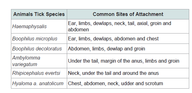
Different tick species have different locations of attachment. The location can indicate the type of tick. Table 1 shows the sites of attachment of different tick species.
Prevention of Ectoparasites
Rather than waiting until the problem of ectoparasites becomes serious, farmers should maintain a strict preventative regimen to controlling ectoparasites. Conduct a thorough physical evaluation of their animals at least once weekly. Run their hand over each animal’s hair coat, visually inspecting for excessive hair loss, flakes of loose skin, areas of skin irritation, and any crusty lesions or bumps that might indicate infection with an external ectoparasites. Immediately separate and place any animal that shows sign of parasite infection or seems to be unthrifty. This helps to reduce the chances of passing infection on to the rest of their animals. Quarantined animals should not be mixed with the main flock until treatment is complete and the parasite eradicated. Isolate newly introduced animals and treat them for ectoparasites before mixing them with other animals [26].
Practice good sanitation habits; Clean animal houses regularly, seal with cement or mud all cracks in the floor and walls of livestock housing, remove grass/plants around the barn and all litter and discarded wool must be collected and burnt or deposited out of animal contact.
Spray housing with an appropriate pesticide every two weeks if possible. Farmers should also be aware of ways to reduce the number of ticks on pasture rotate the land where livestock graze, avoid pasture which has many ticks as long as possible, Chickens can be kept in places where there are many ticks, for example around watering places, etc.
Status of Ectoparasites in Ethiopia
Reports on ectoparasites in Ethiopia are scanty and if present are also very fragmented. Studies conducted at various localities of the country and tanners report on magnitudes of skin pelts damage due to skin disease especially ectoparasites indicated that domestic animals skin disease are becoming growing threat for animals production and export of skin in Ethiopia.
The main ectoparasites reported in Ethiopia are as follows.
Mage mites
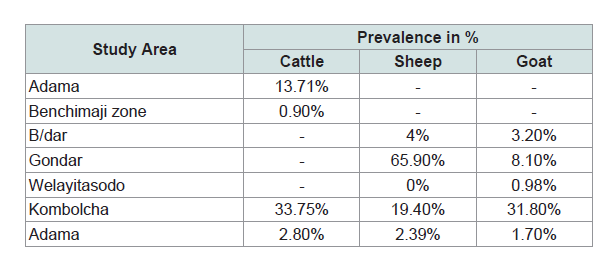
Mange of domestic animals was reported from different area of the country with different magnitude ranges from 0.9% to 65.9% in sheep, goats, cattle and other are indicted in Table 2.
Prevalence of flea infestations in Ethiopia
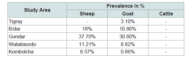
Flea of domestic animals was reported from different area of the country with different magnitude ranges from 8.57% to 37.7% in sheep and 0.66% to 30.6% in goats are indicted in Table 3.
Prevalence of sheep keds (Mallophaga ovinus)
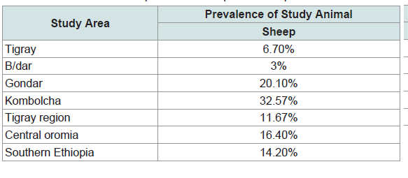
Mallophagus ovis is other ectoparasites that affect the production and productivity of sheep in Ethiopia. However, information on prevalence and distribution of sheep keds are scarce, but examination of sheep in some part of the country indicates that the prevalence of sheep keds ranges from 3% to 32.57%. This was indicted in Table 4 [30,31].
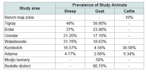
Prevalence of tick infestation
Reports from different area of the country indicate that ticks are also among the ectoparasites affecting domestic animals in Ethiopia. The overall prevalence of ticks infestation ranges from 4.77% to 48% in sheep, 2.68% to 80.7% in goat, and 6.34% to 39.58% in cattle. Table 5 shows that the prevalence of ticks in different part of the country [32,33].
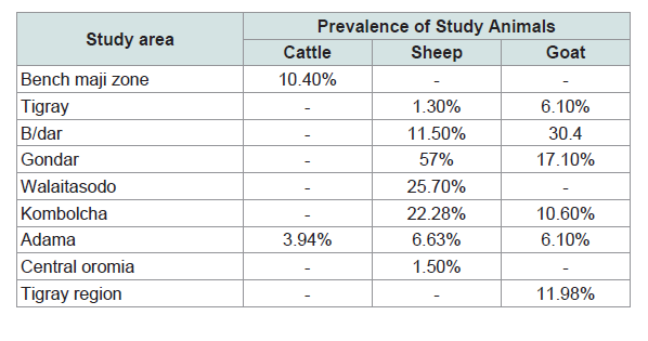
Prevalence of lice infestation
Reports from various part of the country indicate that lice are other ectoparasites of the skin diseases affecting domestic animals in Ethiopia [30]. Total prevalence of louse infestation was reported ranges in cattle from 3.94% to 10.4% in sheep from 1.3% to 57% and in goat it ranges from 6.1% to 30.4%. Totally Table 6 shows the prevalence of louse infestation in different areas of the country.
Conclusion and Recommendation
In Ethiopia the contribution of livestock subsector to the country economy remains marginal compared with the largest livestock potential of the country. This is due to the disease caused by diversified etiology. Epidemiological studies conducted on a diseases caused by ectoparasites of the domestic animals have clearly demonstrated the impact of the problem both on the health of animals and theirperformance. Ectoparasites of the animals are currently a disease of considerable importance in domestic animals production sector as a major cause of down grading and rejection of skin and hide in Ethiopia.
It has been revealed that external parasites are not an outcome of a single determinant, but also consequent to the effects of multiple factors such as poor management, poor plan of nutrition and hygienic conditions. Therefore improving husbandry practices and veterinary services may reduce the level of ectoparasites.
The economic losses by the disease are also the result of reduction in productivity, reproductive performance and death of the affected animals. Moreover, the impact of the disease caused external parasites is severely limiting the performance of the tanning industries, which in turn affect the country’s foreign currency warranted an urgent control intervention.
Based on the above conclusion remarks, the following recommendations are forwarded:
- Further studies should be conducted on the epidemiology of ectoparasites and skin disease in different agro- ecological parts of the country.
- All possible economic losses due to the disease should be assesses at different stages of skin and hide processing including at its early trading stages and in different tanneries.
- Public education should be created on the effect of skin disease on production, productivity, skin/hide quality as well as howto care and handle these products.
- The government, private sectors and veterinarians should work together in order to minimize ectoparasites and their impact.
References
- Wall RL, Shearer D (2001) Veterinary ectoparasites: Biology, pathology and control (2nd edn). Blackwell Science, UK, pp. 304.
- Gross TL, Ihrke PJ, Walder EJ, Affolter VK (2005) Skin disease of the dog and cat: Clinical and histopathologic diagnosis (2nd edn). Blackwell Publishing Science, UK, pp. 11-555.
- González A, Castro DC, González S (2004) Ectoparasitic species from Canis familiaris (Linné) in Buenos Aires province, Argentina. Vet Parasitol 120: 123-129.
- Van den Broek AH, Huntley JF, Halliwell RE, Machell J, Taylor M, et al. (2003) Cutaneous hypersensitivity reactions to psoroptesovis and Der p 1 in sheep previously infested with P. ovis--the sheep scab mite. Vet Immunol Immunopathol 91: 105-117.
- Berriatua E, French NP, Wall R, Smith KE, Morgan KL (1999) Within-flock transmission of sheep scab in naive sheep housed with single infested sheep. Vet Parasitol 83: 277-289.
- Abebayehu T, Endris F, Berhan M, Rahmeto A, Solomon M, et al. (2011) Study on the prevalence of ectoparasite infestation of ruminanats in and around kombolcha and damage to fresh goat pelts and wet blue (pickled) skin at Kombolch Tannary, Northestern Ethiopia. Ethiop Vet J 15: 87-101.
- Hopla CE (1982) Arthropodiasis. CRC Press, Boca Raton, pp. 215-247.
- CSA (Central Statistics Authority) (2004) Prevalence of ectoparistes fauna of ruminants in Ethiopia, Addis Ababa.
- Rahmeto A, Makelesh T, Bekele M, Desie S (2011) Prevalence of small ruminant ectoparasites and associated risk factors in selected districts of tigray region, Ethiopia. Global Veterinaria 7: 433-437.
- Asmare A, Assefa K, Tewodros F (2012) Occurrence of small ruminant ectoparasites in and around Bahir Dar, Northwest Ethiopia. Adv Biol Res 6: 170-176.
- Tewodros F, Fasil W, Mersha C, Malede B (2012) Prevalence of ectoparasites on small ruminants in and around Gondar town. Am Euras J Sci Res 7: 106-111.
- Asnake F, Yacob HT, Hagos A (2013) Ectoparasites of small ruminants in three agro-ecological districts of Southern Ethiopia. Afr J Basic Appl Sci 5: 47-54.
- Leach I (1998) Economic importance of hides, skins and leathers in Ethiopia. Control of sheep and goat skins disease for improved quality of hides and skins MOA/FAO: 21.
- Abegaz M (1999) Quality control and certification in tanning sector. Presented on the occasion of Ethio-Italian industrial partnership meeting in the leather sector/Addis Ababa, Ethiopia. 1227-1247.
- Tewodros F, Mekash, Mersha C (2012) Demodex and sarcoptes mites of cattle: An extravagance for leather industry. Am Euras J Sci Res 7: 131-135.
- Taylor MA, Coop RL, Wall RL ( 2007) Veterinary parasitology (3rd edn). Black well Science, UK, 874.
- Bekele T (2002) Study on seasonal dynamics of tick of ogaden cattle and individual variation in resistance to ticks in Eastern Ethiopia. J Vet Med B Infect Dis Vet Public Health 49: 285-288.
- Hopla CE, Dureden LA, Keirans JE (1994) Ectoparasites and classification. Rev Sci Tech 13: 985-1017.
- Lehmann T (1993) Ectoparasites: Direct impact on host fitness. Parasitol Today 9: 8-13.
- Walker AR (1996) Amblyomma tick feeding in relation to host health. Trop Anim Health Prod 28: S26-S28.
- Lapage G (1968) Veterinary Parasitology (2nd edn) Oliver and Boyd, Edinburgh, London, UK, pp. 634-674.
- Maa TC (1969) A revised checklist and concise host index of Hippoboscidae (Diptera). Pacific Ins Monogr 20: 261-299.
- Leudke AJ, Jochim MM, Bowne JG (1965) Preliminary bluetongue transmissions with the sheep ked Melophagus ovinus (L.). Can J Comp Med Vet Sci 29: 229-231.
- Stacy MD, Angela S, Anna B (2003) Sheep ked Melophagus ovinus. Purdue University, Animal Science Sheep Research and Education Center.
- Durdenl LA, Musser GG (1994) The sucking lice (Insecta, Anoplura) of the world: A taxonomic checklist with records of mammalian hosts and geographical distributions. Bull Am Mus Natur Hist 218: 1-90.
- Desta H (2010) Control of external parasites of sheep and goats. pp. 1-16.
- Pangui LJ (1994) Gale’s des animaux domestiques et méthodes de lutte. Rev Sci Tech Off Int Epiz 13: 1227-1247.
- Richard W, David S (2008) Veterinary ectoparasite: Bilology, pathology and control (2ndedn). Blackwell Science, UK, pp. 1-2.
- Durden LA, Hinkle NC (2009) Fleas (Siphonaptera) in Medical and Veterinary Entomology (2ndedn). Academic Press, San Diego, USA, pp. 115-136.
- Yacob HT, Yalew TA, Dinka AA (2008) Ectoparasite prevalences in sheep and in goats in and around Wolaita Soddo, Southern Ethiopia. Revue Méd Vét 159: 450-454.
- Enquebaher K, Etsay K (2010) Epidemiological study on manage mite, lice and sheep keds of small ruminants in Tigray region, Northern Ethiopia. Ethiop Vet J 14: 51-65.
- Fufa A, Jonsen T, Alemayehu R (2012) Status of tick infestation in small ruminants of Bedelle District, Oromia region, Ethiopia. Trop Anim Health Prod 8: 459-462.
- Tesfaheywet ZS, Simeon HO (2013) Prevalence of ectoparasite infestations of cattle in Bench Maji zone, Southwest Ethiopia. Vet World 6: 291-294.