
Journal of Urology & Nephrology
Download PDF
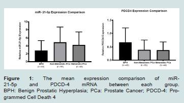 Figure 1: The mean expression comparison of miR-21-5p and PDCD-4 mRNA between each group. BPH: Benign Prostatic Hyperplasia; PCa: Prostate Cancer; PDCD-4: Programmed Cell Death 4
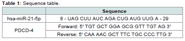 Table 1: Sequence table.
Table 2: Demographic characteristics of recruited participants.
Table 1: Sequence table.
Table 2: Demographic characteristics of recruited participants.
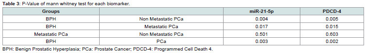 Table 3: P-Value of mann whitney test for each biomarker.
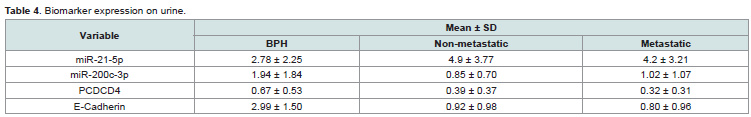 Table 4: Biomarker expression on urine.
Research Article
MicroRNA and messenger RNA As Potential Urinary Biomarkers in Prostate Cancer
Danarto R1, Astuti I2, Umbas R3, and Mubarika Haryana S4*
1Department of Surgery, Universitas Gadjah Mada, Indonesia
2Department of Pharmacology, Universitas GadjahMada, Indonesia
3Departmentof Urology, Universitas Indonesia, Indonesia
4Postgraduate Doctoral Program, UniversitasGadjahMada, Indonesia
*Address for Correspondence: Mubarika Haryana S, Postgraduate Doctoral Program, Faculty of Medicine, Universitas Gadjah Mada, Yogyakarta, Indonesia 55281, Indonesia; E-mail: Sofia.mubarika@gmail.com
Submission: 11 July, 2020;
Accepted: 5 October, 2020;
Published: 9 October, 2020
Copyright: © 2020 Danarto R, et al. This is an open access article distributed under the Creative Commons Attribution License, which permits unrestricted use, distribution, and reproduction in any medium, provided the original work is properly cited.
Abstract
Introduction:
Prostate Cancer (PCa) is the fifth leading cause of death world wide and these condmost common
cancerinmen. Studies to search for new biomarkers,especially with non-invasivemethods, are carried out, one of which is urinary biomarker as an early detection and predictors of PC aprognosis. microRNA (miRNA)
and messenger RNA(mRNA) has proven to have important roles in various oncogenic processes.
Material and methods:
Urine samples collected from 145 patients
were examined,45 patients diagnosed with Benign Prostate Hyperplasia
(BPH) and 100 patients diagnosed with PCa.Urine samples were collected
from each patient and examined in the biomolecular laboratory.The
geneexpression were analyze dusing qPCR analysis using the qPCRCFX 96
thermocycler (Bio-Rad).
Results:
The expression of miR-21-5p was higher in BPH group
compared to PC a groups, both non- metastatic and metastatic, with
p-values of 0.004 and 0.017, respectively. BPH showed the highest mRNA
expression of PDCD-4.
Conclusion:
The overexpression of miR-21-5p shown in this study could
be a potential non-invasive diagnostic tool for patients with PCa. The lower
expression mRNA of PDCD-4 in non-metastatic compared to metastatic
PCa group could be potential prognostic biomarker in PCa.
Keywords
Prostate cancer; Biomarker; miR-21-5p; PDCD-4
Introduction
Prostate Cancer (PCa) isthe fifth leading cause of death
worldwide and the second most common cancer in men. Worldwide,
it was estimated that around 1,276,106 newly diagnosed PCa were
reported in 2018 [1]. The diagnosis of suspected PCa is made when
the abnormality from the Digital Rectal Examination (DRE) and
elevated Prostate Specific Antigen (PSA) present [2]. PSA value of
morethan 4 ng/mlisan indication for prostate biopsy examination
and this border line value hasa positive predictive value of only 37%,
and a negative predictive value of 91% [2]. Therefore, rigorous studies
searching for new biomarkers with non-invasive methods that have
higher specificity than PSA and can be used as an early detectors as
well as prognostic predictors of PCa, have emerged [3].
microRNA (miRNA)is a non-coding RNA molecule which
regulates gene expression and influences both the stability and
the efficiency of target messenger RNA (mRNA) [4]. It has been
proventhat specific miRNA plays a key role in various oncogenic
processes, including angiogenesis, epithelial-mesenchymal transition,
and metastasis. Micro-RNA was suggested to be a potential biomarker
in both serum and urine samples of patients with PCa [3].
miRNA-21 (miR-21) is a specific miRNA which is frequently
up-regulated in cancer and has many targets of tumor suppressors,
such as Phosphatase And Tensin Homolog (PTEN), Programmed
Cell Death 4 (PDCD4), Tropomyosin1 (alpha) (TPM1), Mammary Serine Protease Inhibitor (SERPINB5), and Reversion-
Inducing-Cysteine-Richprotein with Kazal motifs (RECK). It had
been shown that miR-21 has a crucial role in disrupting growth by
inducing apoptosis. miR-21 had used to know the profile CP/CPPS
and clear cell renal cell carcinoma therefore miRNA-21 also used
as a potential marker to know PCa [5,6]. Programmed Cell Death 4
(PDCD-4) is known as a tumor suppressor gene. PDCD-4 decreased
in common tumor entities. The Reduction of PDCD-4 expressions
potential urinary biomarkers in PCa Programmed Cell Death 4
(PDCD-4) is a tumors uppers orgene that has been decreased in
Regulation formany tumor entities. Increased regulation of PDCD-
4 can be found after th einitiation of apoptosis, contrary to the
reduction of PDCD-4expression can contribute tothe anti-apoptotic
nature of cancercells [7]. We aimed to investigate miR-21 and PDCD-
4, expression as potential urinary biomarkers in PCa.
Material and Methods
Sample collection and exosome isolation:
Urine samples collected from 145 patients were examined, 45 patients diagnosed with Benign Prostate Hyperplasia (BPH), and 100 patients diagnosed with PCa. All of the patients participated in this study signed written consent.This study received ethical approval from the Institutional Review Board of Universitas Gadjah Mada (approval number: KE/FK/0449/EC/2019). We collected 15 ml of urine from each patient. The samples were then distributed into fourvials (1.5 mL), and each vial contained 1 mL of the urine sample.
After the centrifugation, the supernatant was extracted and filled into new a vialandkeptina refrigerator at -80°C. The exosomes isolation was conducted using the miRCURY exosome isolationkit (Exiqon, Denmark), by adding 400 uL precipitation buffer B into the vial, and the mixture was then incubated inarefrigerator at 4°C for 60 minutes.
RNA isolation and cDNA synthesis:
The total RNA was extracted using a miRCURYRNA Isolation Kit-Biofluid kit (Exiqon, Denmark). Complementary DNA (cDNA) was synthesized usinga Universal cDNA Synthesiskit (Exiqon, Denmark).
Quantitative polymerase chain reaction (qPCR) and data analysis:
Quantitative PCR was conducted using an ExiLent SYBR Green Master Mixkit (Exiqon, Denmark), primersset (forward and reverse), and diluted cDNA. The qPCR analysis of gene expression was performed using the qPCRCFX96 thermocycler (Bio-Rad). All of the procedures followed the manufacturer’s recommendations, and statistical analyses were performed using the SPSS Version 23 and Graph Pad Prism 7. In this study, statistical significance was setata p-value <0.05 (Table 1 and 2).
Mean age of the samples by BPH, non-metastatic PCa and
metastatic PCa were older than 65 years old. Characteristic median
PSA were appropriate as predisposition lower in BPH rather than
PCa.
The median age of the samples were 65(46-88), 69.5(52-84)
and 67(49-82) for BPH, non-metastatic PCa, and metastatic PCa,
respectively. The median PSA score for BPH, non- metastatic
PCa, and metastatic PCa were 3.8(0.4-20.82), 37.3(2.51-386) and
107.2(23.16-1155) (Table 3,4 and Figure 1).
The expressions of each biomarker were analyzed using Mann
Whitney Test. We found a significant difference between miR-
21-5p expression in the BPH group and PCa, both metastatic and
non-metastatic, withp-values of 0.017 and 0.004, respectively.
Never theless, expression of miR-21-5p between metastatic and
nonmetastasic PCa showed in significant results. BPH group showed
the highest expression of PDCD-4 mRNA. Significant difference was
found between BPH group and PCa group, both metastatic and nonmetastatic.
In this study, miR-21-5p showed potential diagnostic value
todetect PCa. The expression of miR-21-5p, using 1.34 as cut-off
point had 83% sensitivity and 44.4% specificity.
Discussion
Upregulation of urine-based miR-21-5p was significantly higher in patients with PCa than patient with BPH. This study indicated that the overexpression of urine-based miR-21-5p could be apotential non-invasive biomarker for diagnostic aspects of PCa which was aligned with our previous study [8]. This result was supported by previous studies which stated that the overexpression of miR-21in urine and serum samples of PCa patients played an essensial role as a diagnostic and prognostic biomarker [9-13]. PCa proliferation and invasion were significantly decreased by the inhibition of miR-21-5p [14]. Studies conducted by Li T. et al. and Ghorbanmehr, et al. showed increased miR-21-5p expression in urine sample from PCa patients
[15,16]. As one of the best studied miRNAs, miR-21-5p demonstrated its oncogenic activity in most cancers [17]. Additionally, several previous studies suggested that oncomiR miR-21 had been involved in many solid and haematological organ malignancies [18-23]. It has been reported that miR-21-5p was expressed higher in PCa tissue than in normal prostate tissue [24,25]. Melbo-Jorgensen C. et al. showed overexpression of 21-5p in prostate cancer patients undergoing radical prostatectomy [26]. Further more, miR-21-5p, which was shown to contribute to prostate cell transformation and had been associated with cancer initiation, progression, and metastasis [27-29], correlated with stronger PCa cell growth both in vitro and in vivo [30]. In addition, it provided resistance to docetaxelin PC-3 cells, although, knockdown of miR-21 in human cell was sensitive to docetaxel-induced apoptosis [31]. Same study by Kopczynska miRNA had a role in the resistance of prostate cancer with docetaxel and paclitaxel [32]. Study conducted by Porzycki et al. which showed the potential role of miR-21 as a potential diagnostic biomarker, had an analysis done using the ROC curve with the results of the area under the curve of 0.856 [10]. This findings was aligned with the study conducted by Stuopelyte et al. whose area under the curve value was 0.633. The urine specificity of miR-21 was higher than that of PSA (76.22% vs 63.57%) but the sensitivity value was almost the same as
PSA (47.83% vs 52.38%)(33).Our study showed the similar result, where the value of the area under the curve on the ROC miR-21-5p curve is 0.658. Compared with the previous studies, the sensitivity
of miR-21-5p in this study was found to be higher (79%) while the specificity was lower (44.4%).
In this study we found that the expression of PDCD-4mRNA
was lower in PC ag roup than in BPH group. Lowe rexpression
was found in metastatic group compared to non-metastatic group.
This study indicated that lower expression of urine-based PDCD-4
could be a potential non-invasive biomarker for prognostic aspectsof PCa. Several studies showed that the PDCD-4 mRNA was expressed lower in the PCa group compared to BPH. Lower expression was also found inmetastatic group compared to the non-metastatic group. Consistent with previous studies, a significant difference in the PDCD-4 expression was found between PCa and prostatichy perplasia [34]. Down regulation of the PDCD-4 gene in prostate cancer tissue was also found in a study by Fischer N. et al. In PDCD-4 nuclear and cytoplasmic staining, there was a significant decrease
in prostate cancer tissue [25]. Reduced PDCD-4 expression was associated with PCa progression and pathologic features [21,35,36].
Studies conducted in mices, proved that PDCD-4 was a true tumor suppressor [37,38]. A recent study by Aameri, observed localization od PDCD-4 mainly in nuclei epithelial cells in normal but not in prostate cancer specimens [35]. Another study by Zennami, PDCD-4 mRNA and protein level was significantly decreased in higher Gleason
score tumor using 546 patient samples [39]. In a study conducted by
Zennami et al. demonstrated that PDCD4 regulates proliferation, apoptosis and castration resistance in prostate cancer [40]. Matsuhashi et al. found that PDCD-4 protein synthesis was inhibited by miR-21 in prostate cancer. Dong et al. also found that PDCD-4 mRNA expression was decreased in prostate cancer compared to IL-6-inhibited BPH. In this study, it is known that PDCD4 expression in urine can be a diagnostic tool for prostate cancer with a sensitivity value of 90%, but with a specificity of 42.2%. Translation of tumor suppressor gene PDCD-4 is negatively regulated by miR-21(34) [41].
As the target of miR-21 regulationin PC a cells, PDCD-4 expression will bereduced by miR-21, and lower PDCD-4 expression correlates with tumor cell invasion and distant metastasis in PCa. Other studies
showed and rogen stimulation in PCa cellline associated with lower PDCD-4 protein expression, and contribute to miR-21 induced cell growth and castration resistance in PCa [40].
Conclusion
The overexpression of miR-21-5p shown in this study could be a potential non-invasive diagnostic tool for patients with PCa. The lower mRNA expression of PDCD-4 in non-metastatic compared to metastatic PCa group could be potential prognostic biomarker in PCa. Further studies with alarger population are required to investigate the role of miR-21-5p and PDCD-4as biomarkers in PCa. The combination of both durinary based miRNA and mRNA could give potential contribution in management of PCa.
References
Citation
Danarto R, Astuti I, Umbas R, Mubarika Haryana S. MicroRNA and messenger RNA As Potential Urinary Biomarkers in Prostate Cancer. J Urol Nephrol. 2020;7(1): 4.