
Journal of Surgery
Download PDF
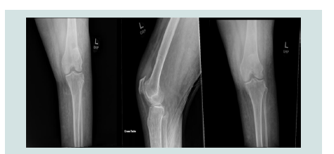 Figure 1: Oblique, lateral, and anteroposterior view of the left knee notable
for effusion surrounding the knee.
Figure 1: Oblique, lateral, and anteroposterior view of the left knee notable
for effusion surrounding the knee.
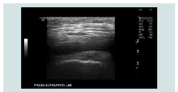 Figure 2: Ultrasound of the left knee showing a small fluid collection of the
medical knee, which appears to be consolidated.
Figure 2: Ultrasound of the left knee showing a small fluid collection of the
medical knee, which appears to be consolidated.
Case Report
A Rare Case of Escherichia Coli Septic Arthritis in a Patient with Klippel-Trenaunay Syndrome
Mele AA*, Santos A, Alaws H, Kumar A, and Hatoum CA
Internal Medicine, Northeast Georgia Medical Center, Gainesville, GA
30501, United States
*Address for Correspondence
Ange Ahoussougbemey Mele, Internal Medicine, Northeast Georgia Medical
Center, Gainesville, GA 30501, United States Email: Ange.Ahoussougbemey@
nghs.com
Submission: 20 February, 2023
Accepted: 25 March, 2023
Published: 29 March, 2023
Copyright: © 2023 Mele AA, et al. Powell BS, et al. This is an open
access article distributed under the Creative Commons Attribution License,
which permits unrestricted use, distribution, and reproduction in any
medium, provided the original work is properly cited.
Abstract
Background:
Septic arthritis, also known as infectious arthritis,
results from an acute invasion of the joint space by microorganisms
that release endotoxins and trigger cytokine release and neutrophil
infiltration. This invasion may happen through the hematogenous
spread, contiguous spread from another locus of infection, or
direct inoculation to a joint. Other causes include iatrogenic from
arthrocentesis or arthroscopy. Bacteria, Mycobacterium, and fungi
are the most common culprits. Patients typically present with joint
pain, swelling, and fever. The condition is associated with increased
morbidity and mortality and thus requires prompt diagnosis and
treatment.
Case Report:
We reported the case of a 55-year-old female with a
past medical history of Klippel-Trenaunay Syndrome (KTS), Escherichia
Coli (E. Coli) bacteremia seven years ago, chronic deep venous
thrombosis, and type 2 diabetes mellitus who presented with chief
complaints of left knee swelling and tenderness. She had Escherichia
Coli septic arthritis. She underwent incision and drainage of the
infected joint and started on four weeks of antibiotics.
Conclusion:
This case is essential as it reports a rare cause of septic
arthritis. Gram-negative bacilli account for only 10% to 15% of all cases
of septic arthritis and are a growing concern. Moreover, it discusses
a practical approach to treating this condition. Patients with KTS are
susceptible to recurrent bouts of cellulitis. However, there is no report
of increasing the risk of septic arthritis, which could have been the
predisposing factor in our patient.
Keywords
Septic arthritis; Escherichia Coli; Gram-negative bacilli;
Orthopedic surgery; Antibiotics selection
Introduction
Septic arthritis is a severe infection of the joint that is associated
with significant morbidity and mortality. Therefore, the clinician
needs to recognize and treat this condition early. E. Coli is a rare
cause of septic arthritis, but its incidence is rapidly increasing. Prompt
orthopedic surgery consultation and a comprehensive antibiotics
selection are essential in tackling this condition.
Case Presentation
A 55-year-old female with a past medical history of KTS;
osteoarthritis; chronic left leg cellulitis on continuous Cephalexin
and Clindamycin for suppression; type 2 diabetes mellitus; chronic
deep vein thrombosis and obesity presented to the hospital with left
knee pain and swelling ongoing for three weeks. She was previously
hospitalizedthree weeks earlier and treated for ten days for left leg and
knee cellulitis. At that time, she received intravenous antibiotics with
Vancomycin and Ceftriaxone.
She stated that her knee continued to be swollen and painful. She
noted that it was excruciating on ambulation, which prompted her to
re-present to the hospital again. She was afebrile on admission, and
the rest of her vitals were within normal limits. Examination revealed knee effusion and significant joint movement pain but did not display
any erythema.
Laboratory work revealed a white blood cell count of 11,300 k/uL
(4.8-10.8 K/uL). The erythrocyte sedimentation rate was 119 mm (0-
30 mm), and the C-reactive protein was 5.1 mg/dL (0.00-0.60 mg/dL).
X-ray imaging showed severe osteoarthritis with an effusion
surrounding the knee (Figure 1). Ultrasound of the left knee
showed a small fluid collection of the medical knee, which appears
to be consolidated (Figure 2). She underwent ultrasound-guided
arthrocentesis with 3 mL of cloudy, bloody fluid obtained. Fluid white
cell count was 58,000 mm3 white blood cells, 95% fluid segs; analysis
of the fluid was negative for crystals.
She underwent an incision, drainage, and irrigation of the left knee
with findings of cloudy-appearing synovial fluid indicating subacute
infection. A culture of the fluid drained from the arthrocentesis grew
E. coli. The culture’s sensitivity showed that the bacteria produced
extended-spectrum beta-lactamase and thus was multi-drug resistant.
Infectious disease decided to treat the patient with intravenous (IV)
Ertapenem 500 mg and IV Daptomycin 12 mg/kg daily for four weeks.
Given that she had a prior history of resistance to E. Coli treatment,
there was suspicion of a gram-positive bacterial component absent in
the culture. This suspicion justifies the addition of Daptomycin to the
antibiotic regimen.
Discussion
In this case report, we have a patient with a complex history of
KTS and past E. Coli bacteremia who came to the ED (Emergency
Department) due to left knee swelling and tenderness which would
later result in E. Coli septic arthritis. Additionally, this patient
received chronic antimicrobial suppressive therapy with clindamycin
and cefalexin for chronic cellulitis. E Coli septic arthritis is a rarity
in the grand scheme of infectious arthropathy. It occurs in patients
with recent abdominal surgery, chronic immunosuppression,
and rheumatoid arthritis. This patient does not have those noted
comorbidities. However, the patient does have KTS. KTS presents
with lymphatic abnormalities, which can occur superficially in
vascular blebs or lymphangiectasis. These abnormalities can also form
deep lymphatic malformations, leading to organ compression and
disfigurement. These lesions are at increased risk for chronic lymph,
blood leakage, or infectious material translocation. Patients with KTS
are susceptible to recurrent bouts of cellulitis. With this information,
these patients with KTS can be chronically at risk for septic arthritis
with various organisms. [1]
As reported by Horowitz et al., it is essential to treat septic
arthritis by initiating antibiotics within the first two days to avoid
complications such as subcartilaginous bone loss, destruction of the
cartilage, and permanent joint dysfunction. The authors postulate
that the most common source of native joint infections is the knee,
hip, shoulder, ankle, elbow, and wrist [2]. Per McBride et al., septic
arthritis has an incidence of 4 to 29 cases per 100,000 person-years.
Patients at higher risk are usually elderly, of lower socioeconomic
status, and immunocompromised. [3].
Lieber et al. report that septic arthritis arises via contiguous, direct
inoculation and hematogenous spread. Indeed, contiguous spread
takes place with a skin infection and cutaneous ulceration. Direct
inoculation happens with a previous intra-articular injection when
a prosthetic joint is placed within the past two years, recent joint
surgery. Hematogenous spread is often seen in Diabetic Mellitus and
HIV infection, use of immunosuppressive medications, intravenous
drug abuse, osteoarthritis, other causes of sepsis, prosthetic joint more
than two years, rheumatoid arthritis, and sexual activity (gonococcal
arthritis). Other risk factors include age older than 80 and smoking
[4].
Visser et al. report that Staphylococcus aureus is the most
common culprit of septic arthritis. Streptococcus pneumonia is the
less prevalent organism but still a leading culprit of adult infection.
Salmonella occurs in sickle cell patients and Pseudomonas in trauma and puncture wound patients. Neisseria gonorrhea is often the most
common cause of acute mono arthritis in sexually active patients.
Fungal and mycobacterial organisms often present subtly but with
harmful effects, making their detection challenging. Usually, a
synovial fluid acid-fast smear is negative; however, in 95% of cases, a
synovial biopsy is positive [5].
Per Chui et al., gram-negative septic arthritis is associated with
poorer outcomes. In gram-negative bacillary septic arthritis, the
cure rate is lower, poorer therapeutic results, recurrent infection,
secondary osteomyelitis, flexion contractures, chronic effusions, and
joint ankylosis have been reported. According to the authors, surgical
drainage is the best treatment for septic arthritis [6].
Margaretten et al. Explain that septic arthritis can is diagnosed by
synovial fluid analysis from the suspected joint. This analysis usually
includes culture, crystals analysis, gram stain, and white blood cell
count with differential). We likely have a bacterial source when the
synovial fluid’s white blood cell (WBC) counts are more significant
than 50,000 and 90% neutrophil predominance. Laboratory tests may
help diagnose septic arthritis, usually including a complete blood
count, an erythrocyte sedimentation rate, inflammatory markers
such as c- reactive protein, ESR, and blood cultures [7]. Horowitz et
al., however, report that in prosthetic joint infections, a White Blood
Cell count of 1100 or more in the synovial fluid that contains 64%
neutrophile predominance indicates septic arthritis [3].
According to Hassan et al., a plain radiograph can demonstrate
widened joint spaces, subchondral bony changes, and bulging of
the soft tissues. It should also be noted that a radiograph does not
disqualify a septic arthritis diagnosis. Ultrasonography may help
identify and quantify the joint effusion and assist in needle aspiration.
MRI is sensitive to detect fluid early, and bone scans help evaluate
localized infections of the sacroiliac or hip joint [8].
Hassan et al. further discuss the treatment of septic arthritis.
According to the authors, antibiotic therapy and joint drainage are
the main courses of treatment. It is essential to initiate antibiotics
after joint fluid aspiration. Options for anti-staphylococcal coverage
include Nafcillin, oxacillin, or vancomycin. Vancomycin may be
used for gram-positive organisms if the physician suspects an MRSA
infection. For additional gram-negative coverage, a third-generation
cephalosporin is optimal. Cultures of the infected material can be
used to direct antimicrobial therapy. The orthopedic surgeon should
determine the procedure to drain the affected joint. This procedure
may include an arthrotomy, arthroscopy, or daily needle aspiration.
Therefore, the orthopedic surgeon’s early involvement is essential [8].
According to Hassan et al., nongonococcal septic arthritis can
be treated for two to four weeks, while extended antibiotic therapy
for about six weeks may be crucial in Pseudomonas aeruginosa.
Gonococcal arthritis responds well to intravenous ceftriaxone for
one to two days. Once clinical improvement occurs, the patient
may start oral therapy to complete their regimen. If there is no
improvement in about one and a half months, the patient should have
another arthrocentesis to rule out Lyme disease, fungal infections, or
reactive arthritis. The exclusion of osteomyelitis occurs by imaging
[8]. Momodu et al. recommend against immobilizing the joint,
and patients should promptly start physical therapy to restore joint mobility and prevent muscle loss. Furthermore, patients with infected
prosthetic joints may undergo joint debridement, prosthesis removal,
and replacement of the new joint with cement-containing antibiotics
[9].
Per Margaretten et al. despite antibiotic use, the mortality rate
is as high as 7% to 15% for in-hospital septic arthritis. One-third of
patients have septic arthritis and morbidity; mortality increases with
age and comorbid conditions. While Neisseria infections rarely result
in death, infection by staphylococcus can carry a mortality rate of
more than 50% [7]. It is, therefore, necessary to promptly recognize
and treat septic arthritis.
Conclusion
E Coli septic arthritis, associated with increased morbidity and
mortality, is a rare and increasingly frequent cause of severe septic
arthritis and other gram-negative bacilli bacteria. KTS may be
associated with an increased risk of septic arthritis associated with
recurrent bouts of cellulitis.
References
Citation
Mele AA, Santos A, Alaws H, Kumar A, Hatoum CA. A Rare Case of Escherichia Coli Septic Arthritis in a Patient with Klippel-Trenaunay Syndrome. J Surgery.
2023;11(1): 3.