
Research Article
Comparative in vitro Performance of an ODU 11/12 Dental Explorer and Differential Reflectometry for Detection of Subgingival Dental Calculus
Thomas E. Rams1,2*, Jennifer A. Lopes1, Micah A.Crowley1 and Susan M. Chialastri1
- 1Department of Periodontology and Oral Implantology, Temple University School of Dentistry, Philadelphia, Pennsylvania,USA
- 2Department of Microbiology and Immunology, Temple University
*Address for Correspondence: Thomas E. Rams, Department of Periodontology and Oral Implantology, Temple University School of Dentistry, Philadelphia, Pennsylvania 19140, USA, Tel: (215) 707-2941; Fax (215) 707-4223; E-mail: trams@temple.edu
Citation: Rams TE, Lopes JA, Crowley MA, Chialastri SM. Comparative in vitro Performance of an ODU 11/12 Dental Explorer and Differential Reflectometry for Detection of Subgingival Dental Calculus. J Oral Biol. 2017; 4(2): 5.
Copyright: © 2017 Rams TE, et al. This is an open access article distributed under the Creative Commons Attribution License, which permits unrestricted use, distribution, and reproduction in any medium,provided the original work is properly cited.
Journal of Oral Biology | ISSN: 2377-987X | Volume: 4, Issue: 1
Submission: 17 May, 2017 | Accepted: 30 June, 2017 | Published: 06 July, 2017
Keywords
Dental calculus; in vitro; Diagnosis
Abstract
Background: The ODU 11/12 dental explorer and differential reflectometry have been used for subgingival dental calculus detection, but little or no data is available to validate their ability to reliably discriminate between dental calculus-positive and -negative tooth root surfaces. This study comparatively assessed, using an in vitro typodont model system, the ability of an ODU 11/12 dental explorer and a DetecTar differential reflectometry device to accurately identify subgingival dental calculus on tooth root surfaces.
Materials and Methods: Three typodont models containing a total of 108 subgingival sites on mandibular posterior plastic teeth, of which 57 (52.8%) had artificial dental calculus deposits, were mounted in manikin phantom heads and scored for subgingival calculus by a team of periodontist examiners using an ODU 11/12 dental explorer and a DetecTar differential reflectometry device. Sensitivity, specificity, positive predictive value, negative predictive value, and accuracy (diagnostic effectiveness) of the two subgingival calculus detection methods were calculated and compared
Results: The ODU 11/12 explorer provided higher sensitivity (91.2% versus 75.4%), better negative predictive values (88.9-89.8% versus 75.9%), and higher overall accuracy (diagnostic effectiveness) (85.2-88.9% versus 80.6%) than differential reflectometry for the detection of subgingival calculus on all types of mandibular posterior tooth surfaces. On proximal tooth surfaces, the DetecTar differential reflectometry device had an inferior level of accuracy for subgingival calculus detection (72.2%) as compared to the ODU 11/12 explorer (83.3-85.2%).
Conclusion: The ODU 11/12 dental explorer provided a high level of in vitro discrimination between subgingival calculus-positive and -negative tooth surfaces, and diagnostically performed in vitro better than a DetecTar differential reflectometry device, supporting its continued use in dental licensure examinations and dental practice for detection of subgingival dental calculus. However, before definitive clinical recommendations can be made, additional efficacy studies in vivo of both subgingival dental calculus detection methods must be performed.
Introduction
The presence of subgingival dental calculus is an important risk factor in the pathogenesis of human periodontitis [1], and its removal from diseased tooth root surfaces constitutes a critical clinical endpoint in periodontal therapy [2].
Subgingival calculus is clinically challenging to reliably detect in periodontal pockets. Various types of dental explorers and periodontal probes are tactilely used to feel for subgingival surface irregularities in lieu of direct visual inspection of tooth roots. High false negative diagnostic findings have been reported with conventional manual explorer and periodontal probe detection of subgingival calculus [3]. After the completion of periodontal root instrumentation, 77.4% of root surfaces still positive for subgingival calculus were wrongly judged as calculus-negative by periodontists using a CP-8 periodontal probe and a G-2 subgingival explorer [3]. Another study where two clinical examiners scored the same periodontal sites for subgingival calculus with an extended 3CH cowhorn explorer after the completion of periodontal root instrumentation found poor agreement between their assessments, with an overall joint agreement of only 21.8% [4].
The Old Dominion University (ODU) 11/12 dental explorer (Hu-Friedy Manufacturing Company, Chicago, IL) is presently used to identify subgingival dental calculus in dental licensure examinations conducted by 4 of the 5 regional clinical testing agencies in the United States, including the Council of Interstate Testing Agencies, the Central Regional Dental Testing Services, the Commission for Dental Competency Assessments (formerly the Northeast Regional Board of Dental Examiners), and the Western Regional Examining Board [5-9]. However, no data is presently available validating the ability of the ODU 11/12 explorer to reliably discriminate between dental calculus-positive and -negative tooth root surfaces. It is not presently known if the ODU 11/12 explorer demonstrates a high or poor level of accuracy in detecting subgingival dental calculus.
Another approach to subgingival calculus detection involves use of differential reflectometry. The DetecTar instrument (NEKS Technologies, Laval, Quebec, Canada) employs a light emitting diode (LED) to deliver red light at a wavelength of 635 nm through a flexible optical fiber extending to the tip of a periodontal probelike hand piece. The optical fiber then collects light reflected back from illuminated tooth surfaces, from which the optical signature of dental calculus is differentiated from that of pristine root surfaces via its unique red light spectra [10]. In 2003, the Food and Drug Administration granted approval for marketing in the United States of the device for detecting subgingival calculus in humans.
However, only a limited amount of research has been conducted to date on the DetecTar device. In vitro, DetecTar correctly differentiated between calculus-positive and -negative root surfaces on 20 extracted teeth, and detected calculus deposits as small as 0.1 mm in size [11]. DetecTar exhibited higher in vitro accuracy and reproducibility than a Williams periodontal probe for detection of subgingival dental calculus on 30 extracted human teeth mounted into manikin heads with silicone gingiva [12]. In vivo, 89% of DetecTar instrument-positive root surfaces were found to have calculus present, whereas 90% of DetecTar-negative root surfaces were calculus-free, in microscopic examinations conducted on 96 human teeth after extraction [13]. Additionally, 83% of tooth root surfaces scored as DetecTar-negative after the completion of periodontal scaling and root planning were found to be calculus-free in post-extraction microscopic examinations [13].
In light of the limited amount of research data on these two approaches to subgingival calculus detection, the purpose of this study was to comparatively assess, using an in vitro typodont model system, the ability of an ODU 11/12 dental explorer and the DetecTar differential reflectometry device to accurately identify subgingival dental calculus on tooth root surfaces.
Materials and Methods
In vitro typodont model system
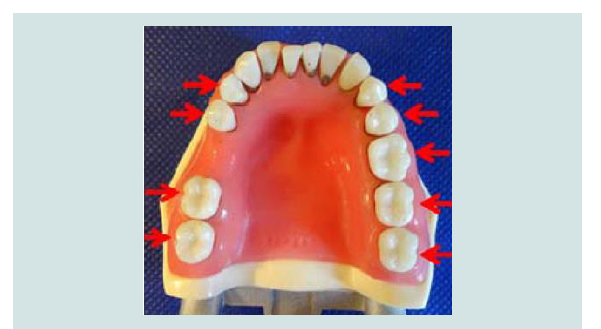
A total of 108 subgingival sites on mandibular posterior plastic teeth, of which 57 (52.8%) had artificial dental calculus deposits, were mounted into 3 typodont models of the human oral cavity, in which plastic teeth are surrounded by anatomically-accurate pink silicone gingiva and palatal soft tissues (Kilgore model P15DP-TR56C GSF, Kilgore International Inc., Coldwater, MI). Mandibular posterior teeth (9 premolar and molar teeth per typodont) alone were used to standardize access/visualization for examiners and provide more challenging in vitro conditions than typically found on anterior teeth (Figures 1). Plastic teeth with dental calculus deposits were randomly distributed throughout the 3 typodont models, and positioned so that the dental calculus was located subgingival to the coronal edge of the gingival soft tissues. The artificial dental calculus deposits were mostly in the form of ledges or rings on calculus-positive root surfaces similar to untreated periodontitis patients. Each typodont was attached to a rubber manikin phantom head with simulated soft tissue mouth shrouds. Defibrinated donor sheep blood (Quad Five, Ryegate, MT) was irrigated into subgingival and interproximal areas around the typodont teeth to simulate the presence of gingival inflammation. Artificial saliva (Aquoral Spray, Bi-Coastal Pharmaceutical Corp., Red Bank, NJ) was applied onto supragingival surfaces to further simulate normal human oral cavity conditions.
ODU 11/12 dental explorer evaluations 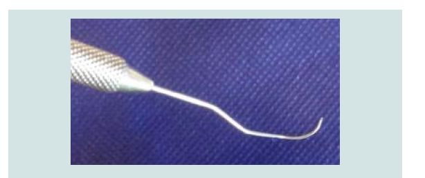 Figure 2: Curved tip of ODU 11/12 dental explorer.
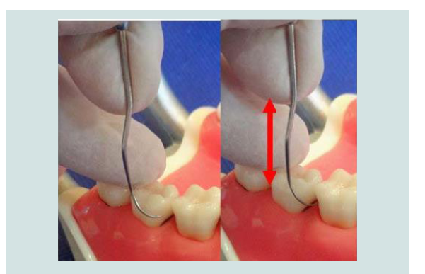
The mandibular posterior tooth surfaces were evaluated for subgingival calculus with an ODU 11/12 dental explorer in duplicate by a primary examiner (author Rams TE), a board-certified periodontist with 30 years of clinical specialty experience. An approximately 20 minute period separated initial and duplicate assessments of each typodont. A secondary examiner (author Chialastri SM), a periodontist with 45 years of dental hygiene/periodontics clinical experience, also scored all test tooth surfaces once. Both examiners were blind to the distribution of subgingival calculus on the mandibular posterior tooth surfaces, and were previously calibrated with a standardized technique for subgingival calculus detection. The ODU 11/12 explorer was employed as previously described [13], with the instrument held parallel to the long axis of teeth, and the side of the curved tip adapted to the contour of tooth surfaces (Figure 2). Subgingival calculus was tactically detected as a jump, bump, or rough surface irregularity characteristic of dental calculus [9], occurring as the explorer was gently moved in repeated apical-coronal vertical and oblique strokes parallel to tooth root surfaces (Figure 3).
ODU 11/12 dental explorer evaluations 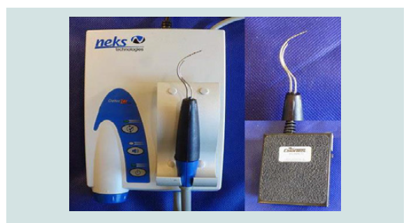 Figure 4: DetecTar differential reflectometry device comprised of an internal computer housed inside a unit base (left) connected to an optical probe embedded within a periodontal probe-like handpiece (upper right),and activated by depressing a foot-switch (lower right).
The mandibular posterior tooth surfaces were also evaluated with a DetecTar differential reflectometry device (NEKS Technologies, Laval, Quebec, Canada) (Figure 4), which was calibrated per manufacturer instructions prior to each use. Emission of a sustained audible signal tone from the DetecTar device following entry of its optical fiber tip into typodont periodontal pockets indicated presence of subgingival dental calculus. Duplicate DetecTar readings were performed by a single periodontist examiner (author Lopes JA), who was blind to the distribution of subgingival calculus on the mandibular posterior tooth surfaces, and previously calibrated with a standardized technique for subgingival calculus detection identical to the other two examiners. An approximately 20 minute period separated initial and duplicate assessments of each typodont with the DetecTar device. In a pilot study, the DetecTar device identified artificial dental calculus deposits on typodont teeth equal to that found with human dental calculus on root surfaces of extracted natural teeth, and the plastic surfaces of typodont teeth were not erroneously identified as dental calculus (data not shown).
Data analysis
Using 2x2 contingency table analysis, the number of true positive (TP), false positive (FP), false negative (FN), and true negative (TN) diagnostic responses with the ODU 11/12 explorer and DetecTar device were each tabulated relative to the in vitro presence or absence of subgingival calculus on mandibular posterior tooth surfaces. From this, the diagnostic performance of each method was determined with calculation of their sensitivity, specificity, positive predictive value, negative predictive value, and accuracy (diagnostic effectiveness) for in vitro detection of subgingival calculus [14].
Sensitivity was defined as the probability that the ODU 11/12 explorer or DetecTar device was diagnostically positive when subgingival calculus was present (true positive rate), and was calculated as TP/TP+FN. Specificity was the probability that the ODU 11/12 explorer or DetecTar device was diagnostically negative when subgingival calculus was absent (true negative rate), and was calculated as TN/FP+TN. Positive predictive value was the probability that subgingival calculus was present when the ODU 11/12 explorer or DetecTar device was diagnostically positive, and was calculated as TP/TP+FP. Negative predictive value was the probability that subgingival calculus was absent when the ODU 11/12 explorer or DetecTar device was diagnostically negative, and was calculated as TN/FN+TN. Accuracy (diagnostic effectiveness) was defined as the proportion of mandibular posterior tooth surfaces correctly categorized as subgingival calculus-positive or -negative by the ODU 11/12 explorer or DetecTar device, and was calculated as (TP+TN)/(TP+TN+FP+FN)[14].
Kappa analysis was performed to quantify agreement beyond chance for the reproducibility of duplicate scoring for subgingival dental calculus with either the ODU 11/12 explorer or DetecTar device [15], and also for the level of agreement between the primary and secondary examiners using the ODU 11/12 explorer. Kappa values between 0.40 and 0.75 were considered to represent fair to good agreement, with those >0.75 indicating excellent agreement.
The PC-based STATA/SE 14.2 for Windows (StataCorp PL,College Station, TX USA) 64-bit statistical software package was used in the data analysis.
Results
For the ODU 11/12 explorer, a good level of reproducibility (kappa=0.62) was found in duplicate scoring of mandibular posterior subgingival surfaces for dental calculus by the primary examiner, and good agreement (kappa= 0.62) was found between the primary and secondary examiners in subgingival calculus detection (data not shown). For the DetecTar device, only a fair level of reproducibility (kappa=0.42) was found in duplicate subgingival calculus scoring of mandibular posterior tooth surfaces by the single periodontist examiner (data not shown).
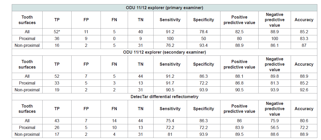
Table 1 presents the in vitro diagnostic performance of the ODU 11/12 explorer and DetecTar differential reflectometry device for detection of subgingival calculus on the 108 mandibular posterior tooth surfaces. The ODU 11/12 explorer provided higher sensitivity (91.2% versus 75.4%), better negative predictive values (88.9-89.8% versus 75.9%), and higher overall accuracy (diagnostic effectiveness) (85.2-88.9% versus 80.6%) than the differential reflectometry device for the detection of subgingival calculus on all types of mandibular posterior tooth surfaces. On proximal (mesial and distal) mandibular posterior tooth surfaces, the DetecTar device yielded an inferior level of accuracy for subgingival calculus detection (72.2%) as compared to the ODU 11/12 explorer (83.3-85.2%).
Discussion
The present in vitro study findings demonstrate that the ODU 11/12 explorer provides reliable identification of subgingival calculus. The instrument yielded reproducible diagnostic assessments, and high levels of sensitivity (91.2%) and accuracy (85.2-88.9%) for subgingival calculus detection, when used by experienced periodontists. The ODU 11/12 dental explorer offers a rapid, reliable, inexpensive, and reproducible method for detecting subgingival dental calculus on tooth root surfaces. The present study data provide additional in vitro validation supporting the continued use of the ODU 11/12 dental explorer for detection of subgingival dental calculus on dental licensure examinations and in clinical practice.
Surprisingly little research has been reported to date on the efficacy of the ODU 11/12 explorer in detecting subgingival calculus. Previous in vitro and in vivo studies have assessed the inter-and intra-rater reliability of clinicians in ODU 11/12 explorer detection of subgingival calculus [16-18]. An in vivo study, using a periodontal endoscope to directly visualize subgingival surfaces, found prior to treatment that only 6.9% of 188 subgingival calculus-positive periodontitis sites were wrongly scored as subgingival calculus-free with the ODU 11/12 explorer [19]. However, after non-surgical root debridement, nearly three-fourths of root surfaces with residual subgingival calculus were erroneously scored as free of subgingival calculus by the ODU 11/12 explorer, highlighting the difficulty of using the instrument to detect small-sized subgingival calculus deposits [19]. No clinical studies to date have assessed the in vivo ability of the ODU 11/12 explorer to detect subgingival calculus on teeth which are subsequently extracted and microscopically examined to determine the true occurrence of subgingival calculus deposits.
In comparison to the ODU 11/12 explorer, scoring of subgingival calculus with the DetecTar differential reflectometry device was less reproducible, and its accuracy on proximal mandibular posterior tooth surfaces was markedly lower. This may be due in part to the design characteristics of the ODU 11/12 explorer, which has a flexible and fine-pointed tip, a curved working end similar to a Columbia 13/14 curette, and a contra-angled shank similar to a Gracey 11/12 curette, which all enhance the instrument’s access into posterior interproximal periodontal pockets [5]. The decreased accuracy of the DetecTar device may also in part be due to a low angulation occurring in tight interproximal areas between the DetecTar probe tip and examined proximal mandibular posterior tooth surfaces, which markedly reduces the dental calculus detection efficacy of the instrument [11]. The diminished diagnostic performance of the DetecTar device on proximal root surfaces, as compared to buccal and lingual areas, is troubling, since deep periodontal pockets are mostly located at interproximal dentition sites [20], where subgingival dental calculus detection is particularly critical to assessing outcomes of periodontal mechanical instrumentation.
The present in vitro study data have several limitations. Artificial dental calculus on plastic typodont teeth was evaluated in vitro instead of human dental calculus in vivo on natural teeth. The amount of dental calculus on the typodont teeth was not quantified and related to its ability to be detected by the ODU 11/12 explorer or DetecTar device. All assessments were made on tooth surfaces devoid of furcation involvements which are frequently found clinically on mandibular posterior teeth with periodontitis [20]. Rubber manikin phantom heads were used, which fail to capture the more challenging intraoral environmental conditions and wide range of other influencing factors clinically encountered on human patients. Only specialists in trained periodontics served as examiners, and those evaluating the ODU 11/12 explorer had 30-45 years of clinical periodontal experience. Since different examiners tested the two instrument/devices, it is possible that the performance differences between the ODU 11/12 explorer and DetecTar device may in part be due to varying levels of examiner error. It is not known whether general dentists, dental hygienists, or less experienced periodontists would attain the same level of diagnostic performance with the two subgingival calculus detection methods as was found with the present study examiners.
Conclusion
The ODU 11/12 dental explorer provided a high level of discrimination between subgingival dental calculus-positive and -negative mandibular posterior tooth surfaces in a typodont model system with experienced periodontist examiners. Based on these in vitro findings, continued use of the ODU 11/12 explorer in dental licensure examinations and dental practice for detection of subgingival dental calculus is supported. In contrast, the DetecTar differential reflectometry device demonstrated lower in vitro accuracy in subgingival calculus detection, particularly on proximal mandibular posterior tooth surfaces, as compared to the ODU 11/12 explorer, and revealed only a fair level of reproducibility in duplicate instrument assessments. Since the overall in vitro performance of the DetecTar device appears to be inferior to that of an ODU 11/12 explorer, its routine clinical utilization in dental practice is not advised based on these findings. However, before definitive clinical recommendations can be made, additional efficacy studies in vivo of both subgingival dental calculus detection methods must be performed.
References
- Mandel ID, Gaffar A (1986) Calculus revisited: a review. J Clin Periodontol 13: 249-257.
- Meissner G, Kocher T (2011) Calculus-detection technologies and their clinical application. Periodontol 55: 189-204.
- Sherman PR, Hutchens LH Jr, Jewson LG, Moriarty JM, Greco GW, et al. (1990) The effectiveness of subgingival scaling and root planing. I. Clinical detection of residual calculus. J Periodontol 61: 3-8.
- Pippin DJ, Feil P (1992) Interrater agreement on subgingival calculus detection following scaling. J Dent Educ 56: 322-326.
- Huennekens S, Daniel SJ (1992) Task analysis of the ODU 11/12 explorer. J Dent Hyg 66: 24-26.
- American Board of Dental Examiners (2017) Patient-based dental examination. Periodontal scaling and restorative sections, 2017 candidate manual, administered by Council of Interstate Testing Agencies, Inc. Council of Interstate Testing Agencies, Inc., Cary, NC, pp. 35.
- Central Regional Dental Testing Services, Inc. (2017) Dental examination candidate manual. Class of 2017, Topeka, KS, USA, pp. 45.
- American Board of Dental Examiners (2017) Patient-based dental examination: restorative and periodontal procedures, 2017 candidate manual, administered by the commission on dental competency assessments. Linthicum, MD, USA, pp. 55.
- Western Regional Examining Board (2017) 2017 dental exam candidate guide. A National Dental and Dental Hygiene Testing Agency, Phoenix, AZ, USA, pp. 75.
- Krause F, Braun A (2004) Detectar™: a novel LED-based optical probe for subgingival calculus detection. Periodontal Pract Today 1: 81-84.
- Krause F, Braun A, Jepsen S, Frentzen M (2005) Detection of subgingival calculus with a novel LED-based optical probe. J Periodontol 76: 1202-1206.
- Shakibaie F, Walsh LJ (2012) Differential reflectometry versus tactile sense detection of subgingival calculus in dentistry. J Biomed Opt 17: 106017.
- Kasaj A, Moschos I, Röhrig B, Willershausen B (2008) The effectiveness of a novel optical probe in subgingival calculus detection. Int J Dent Hyg 6: 143-147.
- Shaikh SA (2011) Measures derived from a 2 x 2 table for an accuracy of a diagnostic test. J Biom Biostat 2: 5.
- Hunt RJ (1986) Percent agreement, Pearson's correlation, and kappa as measures of inter-examiner reliability. J Dent Res 65: 128-130.
- Garland KV, Newell KJ (2009) Dental hygiene faculty calibration in the evaluation of calculus detection. J Dent Educ 73: 383-389.
- Partido BB, Jones AA, English DL, Nguyen CA, Jacks ME (2015) Calculus detection calibration among dental hygiene faculty members utilizing dental endoscopy: a pilot study. J Dent Educ 79: 124-132.
- Santiago LJ, Freudenthal JJ, Peterson T, Bowen DM (2016) Dental hygiene faculty calibration using two accepted standards for calculus detection: a pilot study. J Dent Educ 80: 975-982.
- Osborn JB, Lenton PA, Lunos SA, Blue CM (2014) Endoscopic vs. tactile evaluation of subgingival calculus. J Dent Hyg 88: 229-236.
- Albandar JM, Brunelle JA, Kingman A (1999) Destructive periodontal disease in adults 30 years of age and older in the United States, 1988-1994. J Periodontol 70: 13-29.