
Journal of Forensic Investigation
Download PDF
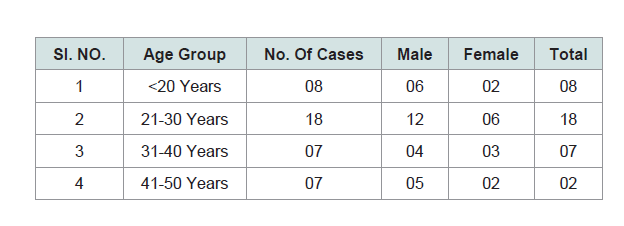 Table 1: Age and Sex distribution of the cases.
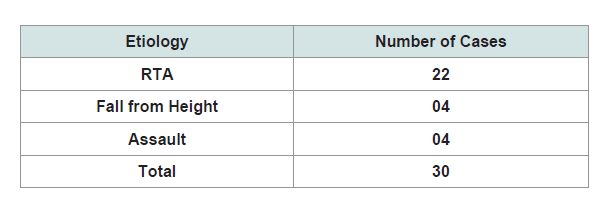 Table 2: Etiology of head injury.
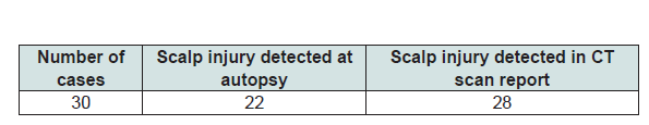 Table 3: Comparison of scalp injury as in Autopsy and CT scan.
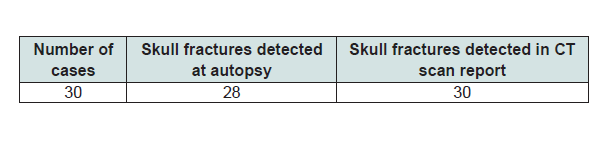 Table 4: Comparison of Skull fractures as in Autopsy and CT scan.
Research Article
A Retrospective Comparison of CT scan Findings and Autopsy Findings in Fatal Head Injury Cases
Vijay Kumar AG, Shivaramu MG, Kumar U, Vinay J* and Somshekar S
Department of Forensic Medicine & Toxicology, Adichunchanagiri
University, India
*Address for Correspondence: Vinay J, Department of Forensic Medicine & Toxicology, Adichunchanagiri Institute of Medical Sciences, B G Nagara, Nagamangala Taluk, Mandya, Karnataka State, India, Phone: 9916735739; E-mail: vijay.fmt@rediffmail.com
Submission: 24 September, 2019;
Accepted: 29 October, 2019;
Published: 31 October, 2019
Copyright: © 2019 Vijay Kumar AG, et al. This is an open access article
distributed under the Creative Commons Attribution License, which
permits unrestricted use, distribution, and reproduction in any medium,
provided the original work is properly cited.
Abstract
A head injury is any injury that results in trauma to the skull or
brain. The terms traumatic brain injury and head injury are often
used interchangeably in the medical literature. All fatal head injury
cases subjected for medico-legal autopsy to the Dept of Forensic
Medicine, Adichunchanagiri Institute of Medical Sciences, where prior
CT Head scan was taken during hospitalization. In the present study,
the vulnerable age group was those in the 21-30 years (18 cases)
followed by age group of < 20 years (8 cases). In the present study, 26
cases were due to RTA injury and remaining 4 cases were due to fall
and assault respectively. In the present study, Of the 30 cases, scalp
injuries were noted in 22 cases at autopsy where as CT reported scalp
injury in only 28 cases. Of the 30 cases, in 28 cases skull fractures were
observed at autopsy but in 30 cases the same was commented upon
in the CT scan. It was observed that combination of CT scan findings
and autopsy findings is a useful tool for the diagnosis of various kinds
of lesions of head injury and thus helps in formulating better policies.
Keywords
Head injury; Autopsy findings; CT scan report
Introduction
A head injury is any injury that results in trauma to the skull or
brain. The terms traumatic brain injury and head injury are often
used interchangeably in the medical literature [1]. Because head
injuries cover such a broad scope of injuries, there are many causesincluding
accidents, falls, physical assault, or traffic accidents-that
can cause head injuries.
The number of new cases is 1.7 million in the United States each
year, with about 3% of these incidents leading to death. Adults have
head injuries more frequently than any age group resulting from
falls, motor vehicle crashes, colliding or being struck by an object,
or assaults. Children, however, may experience head injuries from
accidental falls or intentional causes (such as being struck or shaken)
leading to hospitalization.
A non-contrast CT of the head should be performed immediately
in all those who have suffered a moderate or severe head injury. A
CT is an imaging technique that allows physicians to see inside
the head without surgery in order to determine if there is internal
bleeding or swelling in the brain [2]. Computed Tomography (CT)
has become the diagnostic modality of choice for head trauma due
to its accuracy, reliability, safety, and wide availability. The changes
in microcirculation, impaired auto-regulation, cerebral edema, and
axonal injury start as soon as head injury occurs and manifest as
clinical, biochemical, and radiological changes [3].
Autopsy is the final procedure of choice for finding out the exact cause of death. In head injuries, diagnosis by clinical and radiological
assessment may not reveal the full extent of injuries. In patients who
succumb to their illness, autopsy may detect the lacunae in clinical
diagnosis and investigation. These autopsy findings are a valuable
source of information. This is a unique opportunity to identify the
exact cause of death. It may be possible to modify the protocol for care
of neurotrauma patients in the prehospital and emergency hospital
setting following this study. That is the main purpose of this study.
Objectives
Comparison of autopsy findings with CT scan findings in fatal
head injury cases.
Methodology
Source of data:
All fatal head injury cases subjected for medico-legal autopsy to
the Dept of Forensic Medicine, Adichunchanagiri Institute of Medical
Sciences, where prior CT Head scan was taken during hospitalization.
Study period:
January to December 2018
Method of collection of data:
All fatal cases of head injury subjected for post mortem
examination where ante mortem CT scan reports were available
were taken up for study. Post mortem examination of each case was
carried out as per the standard procedure mentioned in the “Autopsy
diagnosis and technique”. Further a comparative evaluation of post
mortem findings of the head injuries with that of the CT scan report
were analyzed.
Inclusion criteria:
Fatal head injury cases with ante mortem CT Head scan reports
were included in the study.
Exclusion criteria:
Cases where surgical intervention had led to a gross discrepancy between the CT scan findings and autopsy findings were excluded.
Results
(Table 1) The vulnerable age group was those in the 21-30 years
(18 cases) followed by age group of < 20 years (8 cases).
(Table 2) 26 cases were due to RTA injury and remaining 4 cases
were due to fall and assault respectively.
(Table 3) Of the 30 cases, scalp injuries were noted in 22 cases at
autopsy where as CT reported scalp injury in only 28 cases.
(Table 4) Of the 30 cases, in 28 cases skull fractures were observed
at autopsy but in 30 cases the same was commented upon in the CT
scan.
Discussion
In the present study, the vulnerable age group was those in the
21-30 years (18 cases) followed by age group of < 20 years (8 cases).
According to a study by Mukesh K Goyal, Rajesh Verma, Shiv R
Kochar, Shrikant S Asawa where the maximum number of cases i.e.
56 cases (40%) belonged to the age group 21-40 years, followed by
below 10-year age group which were 30 cases (30.4%). Main cause
of injury was Traffic accident (62%). Among males it is 66% and in
females it is 33%. Leading cause of injury among females was fall from
height. Males 122 (87.1%) outnumbered females 18 (12.8%) [4].
Kelly C. Bordignon, Walter Oleschko Arruda observed in their study that highest frequency of Head Trauma occurred in the 21-30
years (25.1%) age group, followed by the age groups 11-20 (21.6%) and
31-40 (17.5%) One thousand three hundred and six (67.3%) patients
were male and 654 (32.7%) were female (sex ratio M: F=2:1) [4]. In
the present study, 26 cases were due to RTA injury and remaining 4
cases were due to fall and assault respectively.
Observation was made by G Gururaj, Sastry Kolluri where RTA
constituted 62%, fall constituted 22% and assault constituted 10% [6].
In the present study, Of the 30 cases, scalp injuries were noted in
22 cases at autopsy where as CT reported scalp injury in only 28 cases.
Of the 30 cases, in 28 cases skull fractures were observed at autopsy
but in 30 cases the same was commented upon in the CT scan.
In a study done by Mohammad Zafar Equabal, Shameem
Jahan Rizvi, Munawwar Husain, V.K Srivastava, Scalp swelling or
haematoma was observed in 86.3% of the cases and the CT Scan
concurred in all cases. It was also the most common CT finding [7].
Sharma R, Murari A in their study observed that amongst skull
fractures, 76.3% of them was diagnosed in both CT scan and Autopsy;
whereas 23.7% of them remained undiagnosed by CT scan [8].
P. Srinivasa Reddy, B. Manjunatha, B.M. Balaraj observed skull
fracture in 48% of the cases at autopsy whereas the same was observed
in only 38 % of the cases in the CT scan [9].
Arvind Kumar et al in their study observed that 69.63 % cases of
head injury had skull fractures [10].
Conclusion
It was observed that combination of CT scan findings and
autopsy findings is a useful tool for the diagnosis of various kinds of
lesions of head injury and thus helps in formulating better policies.
References
Citation
Vijay Kumar AG, Shivaramu MG, Kumar U, Vinay J, Somshekar S. A Retrospective Comparison of CT scan Findings and Autopsy Findings in
Fatal Head Injury Cases. 2019; 7(1): 2.