
Journal of Forensic Investigation
Download PDF
Research Article
*Address for Correspondence:Rashmi Metgud, Professor and HOD, Oral & Maxillofacial Pathology and Microbiology, Pacific Dental College & Hospital, Debari, Udaipur-313024, India, Tel: +91-9001829425; E-mail: rashmi_metgud@rediffmail.com
Citation: Metgud R, Surbhi, Naik S, Patel S. Odontometrics: A Useful Method for Gender Determination in Udaipur Population. J Forensic Investigation. 2015;3(2): 5.
Copyright © 2015 Metgud R, et al. This is an open access article distributed under the Creative Commons Attribution License, which permits unrestricted use,distribution, and reproduction in any medium, provided the original work is properly cited.
Journal of Forensic Investigation | ISSN: 2330-0396 | Volume: 3, Issue: 2
Submission: 4 November, 2015 | Accepted: 18 December, 2015 | Published: 22 December, 2015
Odontometrics: A Useful Method for Gender Determination in Udaipur PopulationTM Identity Panel
Rashmi Metgud*, Surbhi, Smitha Naik and Shrikant Patel
- Oral & Maxillofacial Pathology and Microbiology, Pacific Dental College & Hospital, Udaipur, India
*Address for Correspondence:Rashmi Metgud, Professor and HOD, Oral & Maxillofacial Pathology and Microbiology, Pacific Dental College & Hospital, Debari, Udaipur-313024, India, Tel: +91-9001829425; E-mail: rashmi_metgud@rediffmail.com
Citation: Metgud R, Surbhi, Naik S, Patel S. Odontometrics: A Useful Method for Gender Determination in Udaipur Population. J Forensic Investigation. 2015;3(2): 5.
Copyright © 2015 Metgud R, et al. This is an open access article distributed under the Creative Commons Attribution License, which permits unrestricted use,distribution, and reproduction in any medium, provided the original work is properly cited.
Journal of Forensic Investigation | ISSN: 2330-0396 | Volume: 3, Issue: 2
Submission: 4 November, 2015 | Accepted: 18 December, 2015 | Published: 22 December, 2015
Abstract
Background: Determining gender is one of the most important parameters in forensic identification. Teeth are an excellent material in living and nonliving populations for anthropological, genetic, odontologic and forensic investigations being hardest and chemically the most stable tissue in the body, selectively preserved and fossilized, thereby providing the best records for evolutionary change. Their durability in the face of fire and bacterial decomposition makes them invaluable for identification. Tooth size standards based on odontometric investigations can be used in age and gender determination.Aim and objective : To estimate gender of individuals of Udaipur population by morphometric analysis of teeth and to eval uate and compare specific permanent tooth and dimension for gender determination by using digital vernier caliper.
Materials and methods: The buccolingual and mesiodistal dimensions of all permanent teeth (except second and third molars) of right side (N=25 males and 25 females) belonging to Udaipur, were recorded from the study casts using digital vernier calipers.
Result and conclusion: Males have larger teeth than females and buccolingual dimension is a better gender predictor than mesiodistal dimension. Maxillary canine is found to be more significant in Udaipur population for sexual dimorphism. To conclude, consideration of buccolingual dimension of teeth of Udaipur population can effectively aid in gender determination when both the dimensions are considered together.
Keywords
Teeth; Buccolingual (BL); Mesiodistal (MD); Dimensions; Maxilla; MandibleAbbreviations
BL: Buccolingual; MD: MesiodistalIntroduction
Determining sex from skeletal remains is of paramount importance for archeological and medicolegal examinations. The gender determination is of significance in cases of damaged or mutilated bodies where only fragments of jaw bones with teeth are found then odontometrics plays an important role in determination of sex [1].Sex differentiation in forensic investigation utilizes craniofacial morphology, tooth dimensions and DNA analysis [2,3]. It has been suggested that odontometrics plays an important role in determination of sex in young individuals where secondary sexual characteristics have not developed [2]. Teeth are known to be the most enduring mineralized tissues in the human body [4]. Teeth are resistant to damage in terms of mechanical, thermal, chemical, physical types, makes them valuable for forensic investigation and research [5].
Sex determination using dental features is primarily based upon the comparison of tooth dimensions in males and females or upon the comparison of frequencies of non-metric dental traits like Carabelli’s trait of upper molars, deflecting wrinkle of the lower first molars, distal accessory ridge of the upper and lower canines or shovelling of the upper central incisors [1].
This is based on the fact that although the morphology of the tooth structure is similar in males and females, the size of the tooth does not necessarily remain the same as the tooth size is determined by cultural, environmental, racial, and genetic factors [6].
There are numerous studies in which differences in male and female odontometric features have been identified [4]. Considering the fact that there are differences in odontometric features in specific populations, even within the same population in the historical and evolutionary context, it is necessary to determine specific population values in order to make identification of an individual possible on the basis of odontometrics [4,7].
Gender dimorphism in tooth size has been studied by anthropologists and odontologists focusing on: Buccolingual and Mesiodistal dimensions of teeth and it is known that gender dimorphic dimensions are only useful if relative to a population [3,7-9]. Gender determination using linear dimensions of maxillary and mandibular teeth among the Udaipur population is lacking in the literature. This gap in literature is what this study intends to fill. The present study is an attempt to present odontometrics as an easy-touse, inexpensive, simple, reliable, additional technique to determine sex in archaeological circumstances without need for complicated statistical software.
The focus of this study was to measure the mesiodistal and buccolingual dimensions of the maxillary and mandibular teeth to find out their dimorphic nature. This study will serve as reference data in forensic and physical anthropology as well as guide to dental practitioners in providing clinical information and education in this part of the world.
The present study aimed to determine the mesiodistal and buccolingual dimensions of all maxillary and mandibular permanent teeth (except second and third molars) of right side in both males and females in adult population of Udaipur. In addition, the study intended to evaluate specific permanent tooth and dimension for gender determination among the population chosen.
Significance of the study
Gender dimorphism in tooth size has been studied by anthropologists and odontologists focusing on buccolingual and mesiodistal dimensions of teeth, it is known that gender dimorphic dimensions are only useful if relative to a population. So, it is necessary to determine specific population values in order to make identification of an individual possible on the basis of odontometrics.
Materials and Methods
The buccolingual and mesiodistal dimensions of all permanent teeth (except second and third molars) of right side (N=25 males and 25 females) with the age range between 18-35 years belonging to Udaipur, were recorded from the study casts using digital vernier calipers.The criteria for each measurement were:
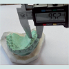
Buccolingual dimension of crown: The greatest distance between buccal and lingual surfaces of crown parallel to the long axis of the tooth (Figures 1 and 2).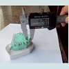
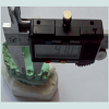
 Mesiodistal dimension of crown: The greatest distance between mesial and distal surfaces of crown perpendicular to the long axis of the tooth (Figures 3 and 4).
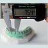
 Periodontally healthy teeth, non carious teeth, teeth with no attrition and intact teeth, satisfactorily aligned maxillary and mandibular teeth, with no spacing or diastema and without crowding, no history or clinical evidence of crown restoration, orthodontic treatment and trauma were considered.
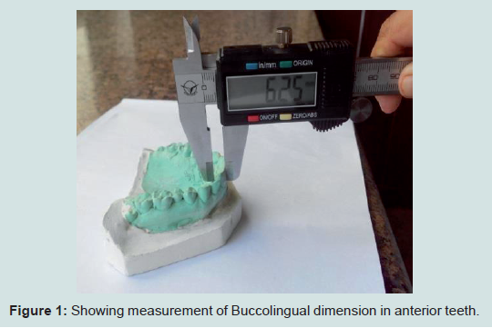
Figure 1: Showing measurement of Buccolingual dimension in anterior teeth.
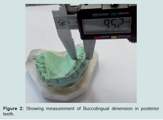
Figure 2: Showing measurement of Buccolingual dimension in posterior teeth.
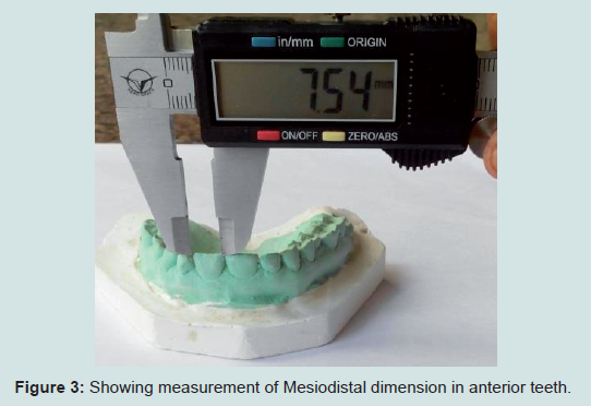
Figure 3: Showing measurement of Mesiodistal dimension in anterior teeth.
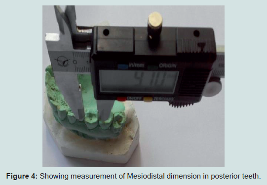
Figure 4: Showing measurement of Mesiodistal dimension in posterior teeth.
The measurements were performed by one person and all values were rounded to two decimal places. In order to assess the reliability of the measurements, intra-observer error was tested. The same measurements were obtained from the original sample at a different time by the same observer to assess intra-observer error. Another observer measured the same selected teeth in order to test the inter-observer error. There was no statistically significant difference between the findings of the two observers.
Statistical analysis
Statistically significant gender dimorphisms in male and female odontometric features were tested by the unpaired t-test. The level of statistical significance was set at p<0.05.
Results
Amount of input DNAIt was observed that the overall comparison of mean values for both BL and MD of both the arches showed highly statistically significant differences for Buccolingual dimensions of maxilla, with p<0.05; measured on the study casts (Table 1).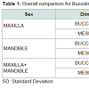 It was evident that on Comparison for Buccolingual dimensions of Maxillary teeth of males and females, statistical significant results were found in maxillary canine and maxillary first molar (Table 2).
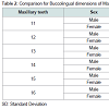 None of the Mandibular teeth showed statistically significant dimorphism on comparison for buccolingual dimensions of Mandibular teeth in males and females (Table 3).
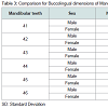 Males showed greater BL dimensions of teeth in comparison to females (Table 4).
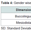 It was evident that on Comparison for Mesiodistal dimensions of Maxillary teeth in males and females, statistically significant results were found in maxillary first molar and maxillary central incisor (Table 5).
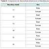 It was evident that on Comparison for Mesiodistal dimensions of Mandibular teeth in males and females, statistically significant results were found in maxillary first molar and maxillary canine (Table 6).
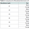
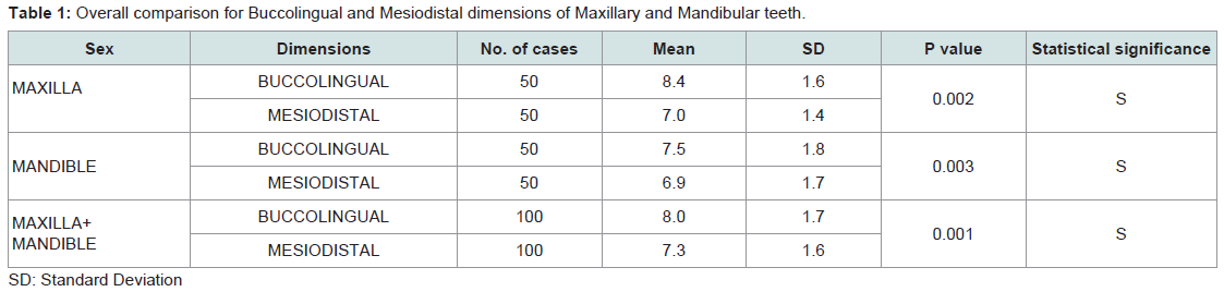
Table 1: Overall comparison for Buccolingual and Mesiodistal dimensions of Maxillary and Mandibular teeth.
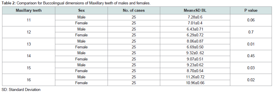
Table 2: Comparison for Buccolingual dimensions of Maxillary teeth of males and females.
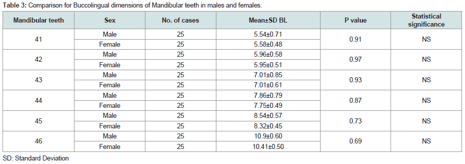
Table 3: Comparison for Buccolingual dimensions of Mandibular teeth in males and females.
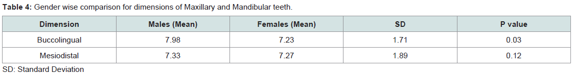
Table 4: Gender wise comparison for dimensions of Maxillary and Mandibular teeth.
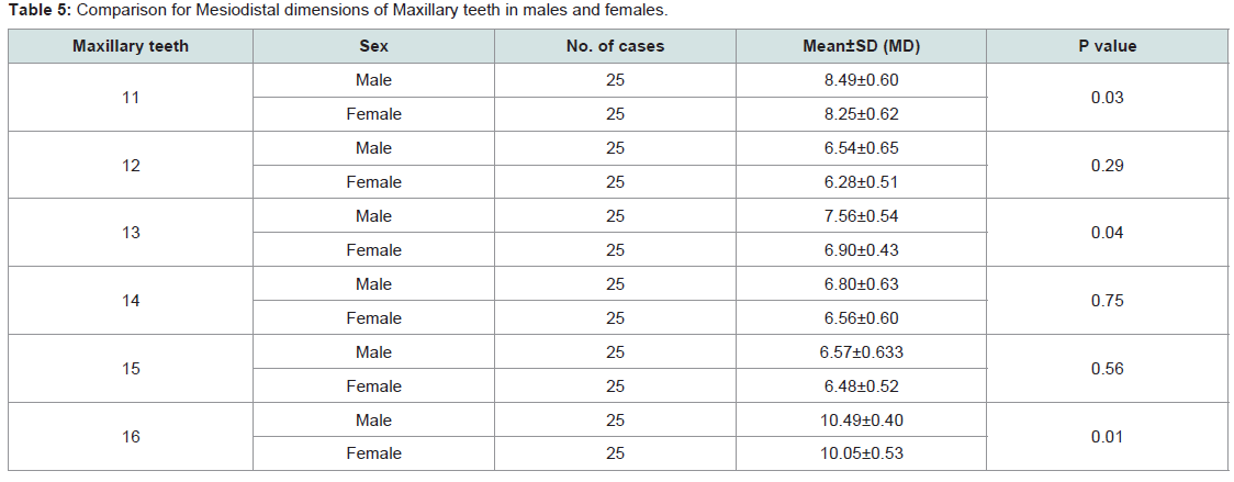
Table 5: Comparison for Mesiodistal dimensions of Maxillary teeth in males and females.
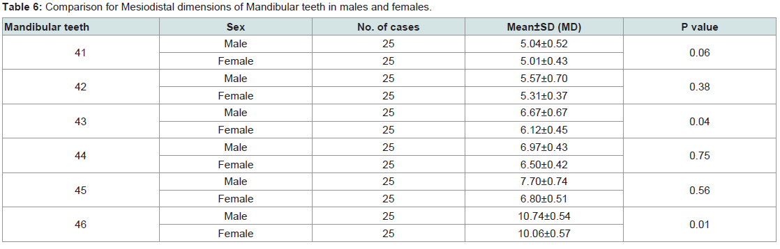
Table 6: Comparison for Mesiodistal dimensions of Mandibular teeth in males and females.
Discussion
Dunn and Dobzhansky have indicated that although all human beings belong to a single species however humans inhabiting different parts of the world are exposed to different environments and are not alike [10]. Various authors like Richardson & Malhotra, Santoro et al., Singh & Goyal have presented literature for tooth dimensions of different populations [11-13] some variations in tooth sizes between gender and among different racial and ethnic groups were observed [14,15].With these considerations, a study was planned to analyze the mesiodistal and buccolingual crown dimensions of the permanent maxillary and mandibular teeth in Udaipur population.
In the maxilla, mean buccolingual crown dimension of canine was larger than first molar which was consistent with findings of other studies (Table 2) [6-8, 16,17]. The mean mesiodistal crown dimension of the first molar was larger than central incisor as reported elsewhere [17].
In the mandible, no statistical significant results were found for buccolingual dimension (Table 3). The mean mesiodistal crown dimension of the first molar was larger than canine as reported elsewhere [6].
In the present study, mean buccolingual crown dimensions of males were found to be larger than those of females for each type of tooth in the maxillary and mandibular arch (Table 4). The results of present studies are inconsistent with other studies, significant differences were observed between sexes for teeth [8,18,19]. This is consistent with Garn et al., Pratibha Rani et al., Eboh, who indicated that the teeth of males were larger than those of females [19,5,20]. Ling et al. also observed sexual dimorphism in permanent teeth [21]. Authors such as (Richardson & Malhotra [11], Singh & Goyal [13]) also demonstrated similar observations [10,12].
Various theories have been given in the literature for this sexual dimorphism:
According to Moss, it is because of the greater thickness of enamel in males due to the long period of amelogenesis as compared to females. However, in females the completion of calcification of the crown occurs earlier in both deciduous and permanent dentition as quoted by de Vito.
Sex chromosomes are also known to cause different effects on tooth size. The ‘Y’ chromosome influences the timing and rate of body development, thus producing slower male maturation, and acts additively and to a greater extent than the ‘X’ chromosome [15].
According to Pratibha et al., ‘Y’ chromosome has a direct effect on tooth size which may be related to a more non-specific effect of hetrerochromatism or cellular activity [5].
Kalia S quoted that according to Townscend, the difference in size has been attributed to differently balanced hormonal production between the sexes consequent to the differentiation of either male or female gonads during the sixth or seventh week of embryogenesis rather than any direct effect of sex chromosome themselves [22].
According to Garn SM et al., there is a low significant correction between sexual dimorphism of teeth and body size and it has been supported by Frayer and Wolpoff [23].
Our results showed that sexual dimorphism was observed for every tooth included in the study between males and females. Besides this, statistically significant dimorphism was exhibited by only permanent maxillary anterior teeth i.e., maxillary canines. Our result of maxillary canine dimorphism was in accordance with other studies who also reported similar findings in their study [6,7,16,17]. Otuyemi and Noar, showed dimorphism in maxillary canines bilaterally [8].
Reason for this dimorphism could be a biologic variation which is a characteristic of life and is attributed to family, genetics and environmental factors [12]. Variation in food resources exploited by different populations has also been explained as one such environmental cause [5]. Previous studies indicate that MD and BL dimensions are more accurate in determining sexual dimorphism [9,24,25]. These can be useful in archeological, odontologic, genetic, and forensic and crime investigations, as ethnicity/race, culture and environment are known to affect odontometrics.
Conclusion
Males had larger teeth than females and buccolingual dimension is a better gender predictor than mesiodistal dimension in the Udaipur study material. Maxillary canine was found to be more significant in Udaipur population for sexual dimorphism. Further studies with larger sample size could provide further evidence of this pattern on gender dimorphism in Udaipur population using mesiodistal and buccolingual dimension.References
- Sonika V, Harshaminder K, Madhushankari GS, Sri Kennath JA (2011) Sexual dimorphism in the permanent maxillary first molar: a study of the Haryana population (India). J Forensic Odontostomatol 29: 37-43.
- Gupta S, Chandra A, Gupta OP, Verma Y, Srivastava S (2014) Establishment of sexual dimorphism in North Indian population by odontometric study of permanent maxillary canine. J Forensic Res 5: 224.
- Lund H, Monstad H (1999) Gender determination by odontometrics in a Swedish population. J Forensic Odontostomatol 17: 30-34.
- Vodanovic M, Demo Z, Njemirovskij V, Keros J, Brkic H (2007) Odontometrics: a useful method for sex determination in an archaeological skeletal population? J Archaeol Sci 34: 905-913.
- Prathibha Rani RM, Mahima VG, Patil K (2009) Bucco-lingual dimension of teeth - An aid in sex determination. J Forensic Dent Sci 1: 88-92.
- Narang RS, Manchanda AS, Malhotra R, Bhatia HS (2014) Sex determination by mandibular canine index and molar odontometrics: A comparative study. Indian J Oral Sci 5:16-20.
- Iscana MY, Kedici PS (2003) Sexual variation in bucco-lingual dimensions in Turkish dentition. Forensic Sci Int 137: 160-164.
- Otuyemi OD, Noar JH (1996) A comparison of crown size dimensions of the permanent teeth in a Nigerian and a British population. Eur J Orthod 18: 623-628.
- Acharya AB, Mainali S (2008) Are dental indexes useful in sex assessment? J Forensic Odontostomatol 26: 53-59.
- Savara BS, Sanin C (1969) A new data acquisition method for measuring dentitions and tests of accuracy. Am J Phys Anthropol 30: 315-318.
- Richardson ER, Malhotra SK (1975) Mesiodistal crown dimension of the permanent dentition of American Negroes. Am J Orthod 68: 157-164.
- Santoro M, Ayoub ME, Pardi VA, Cangialosi TJ (2000) Mesiodistal crown dimensions and tooth size discrepancy of the permanent dentition of Dominican Americans. Angle Orthod 70: 303-307.
- Singh SP, Goyal A (2006) Mesiodistal crown dimensions of the permanent dentition in North Indian children. J Indian Soc Pedod Prev Dent 24: 192-196.
- Ajayi EO, Ajayi YO, Oboro HO, Chukwumah NM (2010) Mesiodistal crown dimensions of the permanent dentition in a Nigerian population. Dental Anthropol 23: 57-60.
- Bunger E, Jindal R, Pathania D, Bunger R (2014) Mesiodistal crown dimensions of the permanent dentition among school going children in Punjab population: an aid in sex determination. Int J Dent Health Sci 1: 13-23.
- Staka G, Bimbashi V (2013) Sexual dimorphism in permanent maxillary canines. Int J Pharm Bio Sci 4: 927-953.
- Lakhanpal M, Gupta N, Rao NC, Vashisth S (2013) Tooth dimension variations as a gender determinant in permanent maxillary teeth. JSM Dent 1: 1014.
- Acharya AB, Mainali S (2008) Sex discrimination potential of buccolingual and mesiodistal tooth dimensions. J Forensic Sci 53: 790-792.
- Garn SM, Cole PE, Wainwright RL, Guire KE (1977) Sex discriminatory effectiveness using combinations of permanent teeth. J Dent Res 56: 697.
- Eboh DE (2012) A dimorphic study of maxillary first molar crown dimensions of Urhobos in Abraka, South-Southern Nigeria. J Morphol Sci 29: 96-100.
- Ling JY, Wong RW (2007) Tooth dimensions of Southern Chinese. Homo 58: 67-73.
- Kalia S (2006) Study of permanent maxillary and mandibular canines and inter-canine arch widths among males and females. Dissertation submitted to the Rajiv Gandhi University of Health Sciences, Bangalore, Karnataka, India.
- Garn SM, Lewis AB, Swindler DR, Kerewsky RS (1967) Genetic control of sexual dimorphism in tooth size. J Dent Res 46: 963-972.
- Narang RS, Manchanda AS, Singh B (2015) Sex assessment by molar odontometrics in North Indian population. J Forensic Dent Sci 7: 54-58.
- Manchanda AS, Narang RS, Kahlon SS, Singh B (2015) Diagonal tooth measurements in sex assessment: A study on North Indian population. J Forensic Dent Sci 7: 126-131.