
Case Report
Coincidence of Tender Facial Swelling and Painful Calf Lesions in a 23 Year Old Woman
Idan Blum*, Majd Khader and Zamir Doron
- Department of Internal Medicine D Barzilai medical Center, University of Beer Sheva, Israel
*Address for Correspondence: Idan Blum, Department of Internal Medicine D Barzilai medical Center, University of Beer Sheva, Ashkelon campus Ashkelon, Israel, E-mail: IBLUM2000@GMAIL.COM
Citation: Blum I, Khader M, Doron Z. Coincidence of Tender Facial Swelling and Painful Calf Lesions in a 23 Year Old Woman. J Clin Med Case Reports. 2019;6(1): 2.
Copyright © 2019 Blum I, et al. This is an open access article distributed under the Creative Commons Attribution License, which permits unrestricted use, distribution, and reproduction in any medium, provided the original work is properly cited.
Journal of Clinical & Medical Case Reports | ISSN: 2332-4120 | Volume: 6, Issue: 1
Submission: 07 January 2019 | Accepted: 06 February 2019 | Published: 08 February 2019
Abstract
A 23 year old female was admitted to Internal Medicine department due to bilateral face swelling of both preauricular area and multiple tender small lesions in the anterior aspect of both calves. A few similar lesions were found on the posterior aspect of her arms. She had a prodrome of two days fever before and upper dysphagia and odynophagia a day before her admission. She reported of mild alcohol consumption and childhood asthma. She did not take any medication prior to her admission except oral contraceptives, which were prescribed for a few weeks, until 4 months ago.
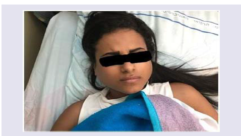
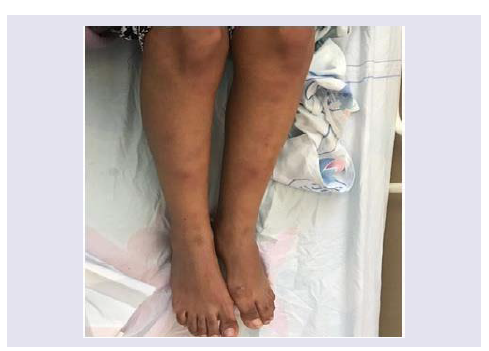
On physical examination, the patient seemed to have bilateral tender swellings of both preauricularareas ,that were painful while opening her mouth (Figure 1). She also had bilateral tender erythematous lesions on both calves and a few similar lesions involving he extensor portion of her arms (Figure 2). The lesion borders were poorly defined, and lesions vary from 2-5 cm. The rest of physicalexamination was within the normal range.
Laboratory examination included elevated CRP (133 mg/dL, norm<0.5 mg/dL). CBC, liver functions and renal functions were all within the normal range. Blood cultures, serology for HAV, HBV, HCV, EBV, CMV and HIV and Mantoux test were negative.
Antinuclear antibody was weak positive (+1), complement levels (c3 & c4) were in normal range and Rheumatoid Factor was negative. Beta HCG was negative (<0.6). Chest X-ray was without pathologic findings.
Keywords
Parotitis; Erythema nodosum; MUMPS; Uncommon case of mumps; Israeli mumps; Multiple tender small lesions; Swelling of preauricular area; Gold standard for EN diagnosis; First coincidence of Mumps causing; Erythema nodosum
Introduction
Mumps is a contagious viral illness that is largely preventable via vaccination. Typically, it begins with a few days of fever, headache, myalgia, fatigue, and anorexia, followed by parotitis; the illness is usually self-limited.
Mumps occurs worldwide and the peak incidence is typically in the late winter to early spring, although sporadic outbreaks occur at any time of year. Mumps occurs most commonly among school-aged children and college-aged young adults and it is rare among infants less than one year of age, who have protection via maternal antibodies.
Mumps is highly infectious and is transmitted by respiratory droplets, direct contact, or fomites. Mumps spreads rapidly among susceptible individuals living in close quarters, such as young adults living in college dormitories. Infection among school-aged children may be associated with further spread to household family members [1].
Clinical Manifestations
Mumps typically begins with a few days of fever, headache, myalgia, fatigue, and anorexia, these manifestations are usually followed by development of salivary gland swelling within 48 hours.
Parotitis occurs most commonly among children between two and nine years of age, tenderness, occasionally associated with earache, typically precedes parotid swelling.
Parotitis may be unilateral or bilateral; initial unilateral involvement is followed by contralateral involvement a few days later in 90 percent of cases. Parotid swelling can last up to 10 days.
On physical examination, parotid swelling may obscure the angle of the mandible, and the orifice of Stensen’s duct is erythematous and enlarged [2].
Laboratory findings include leukopenia with a relative lymphocytosis and an elevated serum amylase concentration.
Asymptomatic infection occurs in 15 to 20 percent of cases and it is more common in adults than in children. Adults who do have symptomatic infection are more likely to have severe manifestations than children with symptomatic infection.
Diagnosis
Physical examination revealed bilateral tender swelling of both parotids and diagnosis of bilateral parotitis, was approved by an Otolaryngologist examination, which ruled out sialolithiasis.
Elevated level of Amylase (532 IU/ml, Norm<90IU/ml) was also compatible with the diagnosis. Buccal scrapping for Mumps was considered and had been sent for mumps PCR, which was negative in hospitalization (sensitivity of 71% from buccal sample) [3].
However serum ELISA examination was positive for mumps, both IgG and IgM (sensitivity of 100%). The parotitis resolved within a few days and the nodules disappeared within 3 weeks with symptomatic NSAID treatment.
Discussion
Erythema Nodosum (EN) is a delayed-type hypersensitivity reaction that most often presents as erythematous, tender nodules on the shins [4]. Common triggers for EN include infection (especially streptococcal pharyngitis, TB, mycoplasma, brucellosis, chlamydia, coccidioidomycosis, histoplasmosis, EBV, Hepatitis virus, Paravaccinia virus), drugs, pregnancy, malignancy, and autoimmune conditions (sarcoid, IBD, bechcet), however many cases are idiopathic. The characteristic histologic finding in EN is a septal panniculitis without vasculitis [5].
The gold standard for EN diagnosis is a histologic examination, however it is not mandatory if diagnosis is clear from physical examination.
Atypical syndromes of sarcoidosis such as Lofgren syndrome and heerfordt-waldenstromsyndrome, were considered.
The second syndrome includes parotitis, fever and facial nerve palsy as a distinct manifestation of sarcoidosis. Normal ACE levels, normal chest x ray and lack of other typical symptoms except EN excluded but did not ruled out completely diagnosis of sarcoidosis, which may manifests as EN and bilateral parotitis, not much different from bechcet, Sjogren syndrome, granulomatosis with polyangitis [6-8].
A careful search in medical literature, including Pubmed, Google and Up To Date did not reveal any reports regarding coincidence of EN and mumps.
We believe that this case report presents the first reported coincidence of Mumpsparotitis causing Erythema Nodosum [9,10].
We believe that this peculiar case may cause physicians to be more aware to such cases and avoid unnecessary antibiotic for the lesions which are uncommon in case of Mumps, as our hospital began to monitor the overwhelming wide spread use of antibiotics.
Conclusion
Since there is little clinical cases suggesting that erythema nodosumcan appear in mumps, we decided to write this clinical case to make clinicians aware that erythema nodosum may appear in mumps parotidis. Thus, we recommend that clinicians must be aware and take into consideration the occurrence of other uncommon symptoms and complains that might appear in mumps, and not only the classic ones.
References
- Porges T, Shafat T, Sagy I, Zeller L, Bartal C, et al. (2018) Clinical, Epidemiological, and Etiological Changes in Erythema Nodosum. Isr Med Assoc J 20: 770-772.
- Gadodia A, Bhalla AS, Sharma R, Thakar A, Parshad R (2011) Bilateral parotid swelling: a radiologic review. Dentomaxillofac Radiol 40: 403-414.
- Zamir CS, Schroeder H, Shoob H, Abramson N, Zentner G (2015)Characteristic of a large mumps outbreak. Hum Vaccin Immunother 11: 1413-1417.
- Garty BZ, Poznanski O (2000) Erythema Nodosum in Israeli children. Isr Med Assoc J 2: 145-146.
- Lührs JP, Krug N, Schürmeyer TH (1997) Swollen parotid gland and lower leg erythema. Presentation of differential diagnosis and therapy based on a case report. Fortschr Med 115: 39-42.
- Fraga RC, Kakizaki P, Valente NYS, Portocarrero LKL, Teixeira MFS, et al. (2017) Do you know this syndrome? Heerfordt-Waldenström syndrome. An Bras Dermatol 92: 571-572.
- CDC guidelines, mumps vaccination.
- Kunz M, Beutel S, Bröcker E (1999) Leucocyte activation in erythema nodosum. Clin Exp Dermatol 24: 396-401.
- Blake T, Manahan M, Rodins K (2014) Erythema nodosum - a review of an uncommon panniculitis. Dermatol Online J 20: 22376.
- Porges T, Shafat T, Sagy I, Zeller L, Bartal C, et al. (2018) Clinical, Epidemiological, and Etiological Changes in Erythema Nodosum. Isr Med Assoc J 20: 770-772.