
Case Report
*Address for Correspondence: Sajazd Ahmad Wani, Department of Paediatric Surgery, SKIMS, Soura, Srinagar 190011, Jammu and Kashmir, India, Tel: +91 9596310531; E-mail: ahmadsajadwani@gmail.com
Citation: Wani SA, Hassan F, Baba AA, Bhat NA, Mufti GN, et al. Acute Perforated Appendix Mimicking Umbilical Hernia: A Rare Case Report. J Clin Med Case Reports. 2016;3(2): 2.
Copyright © 2016 Wani SA, et al. This is an open access article distributed under the Creative Commons Attribution License, which permits unrestricted use,distribution, and reproduction in any medium, provided the original work is properly cited.
Journal of Clinical & Medical Case Reports | ISSN: 2332-4120 | Volume: 3, Issue: 2
Submission: 19 October, 2016 | Accepted: 09 December, 2016 | Published: 15 December, 2016
Acute Perforated Appendix Mimicking Umbilical Hernia: A Rare Case Report
Sajazd Ahmad Wani*, Faheemul Hassan, Aejaz Ahsan Baba, Nisar Ahmad Bhat, Gowher Nazir Mufti and Sheikh Khursheed
- Department of Paediatric Surgery, SKIMS, Soura, Srinagar, Jammu and Kashmir, India
*Address for Correspondence: Sajazd Ahmad Wani, Department of Paediatric Surgery, SKIMS, Soura, Srinagar 190011, Jammu and Kashmir, India, Tel: +91 9596310531; E-mail: ahmadsajadwani@gmail.com
Citation: Wani SA, Hassan F, Baba AA, Bhat NA, Mufti GN, et al. Acute Perforated Appendix Mimicking Umbilical Hernia: A Rare Case Report. J Clin Med Case Reports. 2016;3(2): 2.
Copyright © 2016 Wani SA, et al. This is an open access article distributed under the Creative Commons Attribution License, which permits unrestricted use,distribution, and reproduction in any medium, provided the original work is properly cited.
Journal of Clinical & Medical Case Reports | ISSN: 2332-4120 | Volume: 3, Issue: 2
Submission: 19 October, 2016 | Accepted: 09 December, 2016 | Published: 15 December, 2016
Abstract
The contents of umbilical hernia are usually omentum or bowel. Unexpected contents cause confusion in the diagnosis, especially when inflamed, and may mimic a strangulated umbilical hernia. Only four cases of appendicitis within an umbilical hernia have been reported. We reported a case of perforated appendicitis in to the abdominal wall at umbilicus presenting as strangulated umbilical hernia.Keywords
Perforated appendicitis; Strangulated; Umbilical herniaIntroduction
Acute appendicitis is very common in pediatric patients [1]. The presentation of acute appendicitis is varied due to the different locations of appendix which leads to diagnostic dilemma in a significant number of cases [2]. The diagnosis becomes even more difficult in infants and preverbal children [3]. We hereby share our experience of a patient having perforated appendicitis presenting as umbilical swelling. The purpose of this report is to highlight the necessity of having a suspicion of appendicitis in patients with acute umbilical swellings with unusual presentations.Case History
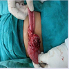
A one year old female patient presented with acute swelling at umbilicus with fever and vomiting since 48 hours. Patient also had developed abdominal distension since last 8 hours. There was no previous history of any swelling at umbilicus. Patient was sick looking and dehydrated. On evaluation patient had tachycardia and fever. Clinical examination revealed a 1 x 2 cm swelling at umbilicus which was tender on palpation. The swelling was indurated and firm and was non-reducible. The cough impulse was absent. Leucocyte counts were raised with neutrophilia. USG abdomen revealed a gut loop adherent to abdominal wall at umbilicus and surrounding fat stranding. There were no signs of obstruction on plain radiograph. Provisional diagnosis of incarcerated umbilical hernia was made and patient was explored after an initial period of resuscitation. On exploration, once the sheath was divided, pus came out. A formal laparotomy was carried and findings revealed mobile caecum and perforated appendix (inferio-laterally on the ileocaecal valve) wrapped by inflamed omentum (Figure 1) with normal rest of the gut. The appendix had perforated into the abdominal wall at umbilicus and had formed an umbilical abscess. The possible aetiology of the perforated appendix in to the umbilicus is mobile caecum. Appendectomy was performed and a fecalith was seen inside the inflamed appendix. Wound was closed back. Postoperatively orals were started on second postoperative day and patient was discharged on third postoperative and is doing well in the follow-up since the surgery.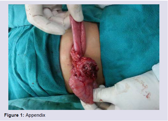
Figure 1: Appendix.
Discussion
Acute appendicitis is a common ailment in pediatric age group. Due to the nonspecific symptoms the presentation and diagnosis may be delayed at times leading to perforation with local or general peritonitis [4]. Perforated appendix has varied presentation depending upon the location of appendix. It can lead to abdominal lump, intraperitoneal and pelvic abscesses and peritonitis. There have been a few reports of finding a perforated appendix in inguinal and femoral hernias, but finding a perforated appendix in umbilical hernia is very rare [5]. In our present case the appendix had perforated through the umbilicus with inflamed omentum surrounding it and the caecum giving it an appearance of incarcerated hernia on ultrasonography. This is possible due to the different anatomical positioning of the appendix and different degrees of gut rotation.Conclusion
High degree of suspicion is needed in such a situations and perforated appendix should be considered especially when the clinical presentation is unusual of an umbilical hernia.References
- Alloo J, Gerstle T, Shilyansky J, Ein SH (2004) Appendicitis in children less than 3 years of age: a 28-year review. Pediatr Surg Int 19: 777-779.
- Marzuillo P, Germani C, Krauss BS, Barbi E (2015) Appendicitis in children less than five years old: A challenge for the general practitioner. World J Clin Pediatr 4: 19-24.
- David OO (2009) Gangrenous retrocolic appendix masquerading as incarcerated umbilical hernia in a 13-month-old boy. J Trop Pediatr 55: 202-204.
- Singh M, Kadian YS, Rattan KN, Jangra B (2014) Complicated appendicitis: analysis of risk factors in children. Afr J Paediatr Surg 11: 109-113.
- Agarwal N, Goyal S, Kumar A, Garg A, Kaur N, et al. (2013) Appendicitis in paraumbilical hernia mimicking strangulation: a case report and review of the literature. Hernia 17: 531-532.