
Journal of Clinical and Investigative Dermatology
Download PDF
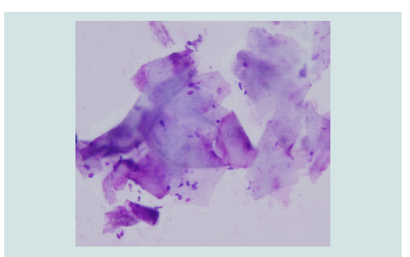 Figure 1: Cytology from Group 1 shows exfoliated superficial squamous cells
and M. furfur.
Figure 1: Cytology from Group 1 shows exfoliated superficial squamous cells
and M. furfur.
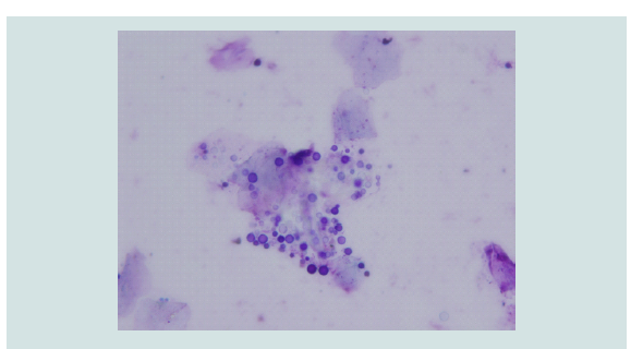 Figure 2: Cytology from Group 1 shows exfoliated superficial squamous cells
and M. globosa.
Figure 2: Cytology from Group 1 shows exfoliated superficial squamous cells
and M. globosa.
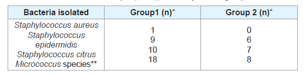 Table 1: Shows the Bacteriology findings of Group 1 (n=40) with dandruff or
seborrheic dermatitis and Group 2 (n=20), a healthy control group.
Table 1: Shows the Bacteriology findings of Group 1 (n=40) with dandruff or
seborrheic dermatitis and Group 2 (n=20), a healthy control group.
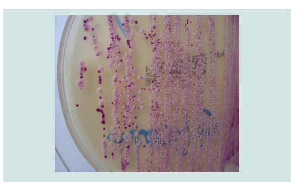 Figure 3: Malassezia furfur colonies were easily distinguishable on CHRO
Magar Malassezia due to their characteristically large pale pink colonies and
M. globosa was small purple colonies.
Figure 3: Malassezia furfur colonies were easily distinguishable on CHRO
Magar Malassezia due to their characteristically large pale pink colonies and
M. globosa was small purple colonies.
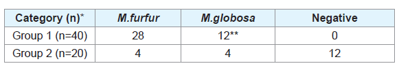 Table 2: Shows the Mycology findings of Group 1 with dandruff or seborrheic
dermatitis and Group 2, the healthy control group.
Table 2: Shows the Mycology findings of Group 1 with dandruff or seborrheic
dermatitis and Group 2, the healthy control group.
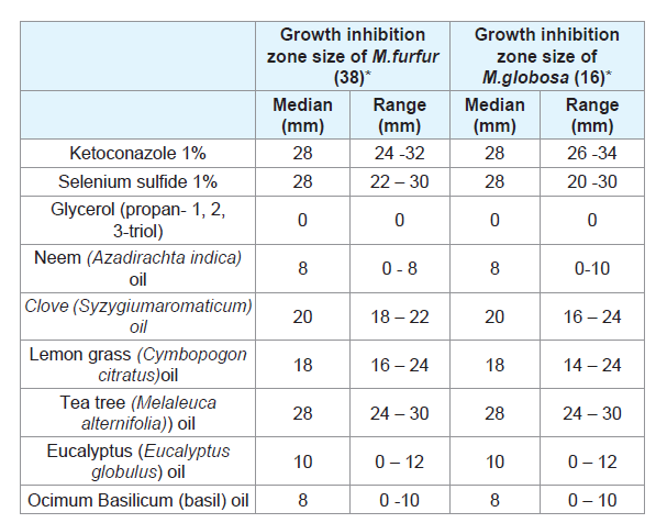 Table 3: Shows the zone of inhibition of herbal oils, azole, and other chemical
agents against M. furfur and M.globosa which were isolated from the scalp of teens.
Table 3: Shows the zone of inhibition of herbal oils, azole, and other chemical
agents against M. furfur and M.globosa which were isolated from the scalp of teens.
Research Article
Role of <i>Malassezia furfur</i> and <i>M. globosa</i> in Dandruff and Seborrheic Dermatitis
Sibi D1*, Silvanose CD2 and Jibin VG3
1Sri Sidhartha Medical College, Tumkuru, Karnataka, India
2Dubai Falcon Hospital, Dubai, UAE
3District Hospital, Bundi, Rajasthan, India
*Address for Correspondence:
Sibi D, Sri Sidhartha Medical College, Tumkuru, Karnataka, India
Email: sdsilvanose@gmail.com
Submission: 12 December, 2022
Accepted: 10 January, 2023
Published: 14 January, 2023
Copyright: © 2023 Sibi D, et al. This is an open access article
distributed under the Creative Commons Attri-bution License,
which permits unrestricted use, distribution, and reproduction in
any medium, provided the original work is properly cited.
Abstract
Dandruff is a common problem in both teens and adults. This study
is to evaluate the role of bacteria and fungi associated with dandruff
and seborrheic dermatitis. Malassezia furfur (70%) was the predominant
isolate, followed by Malassezia globosa (30%) which included mixed
infection (15%) of both M. furfur and M. globosa together adding as
the significant causative agents (p < 0.00001) as compared to healthy
teens. A qualitative in-vitro susceptibility study was performed with
Ketoconazole which showed good in-vitro anti-Malassezia activity
with a greater inhibitory zone, and similar anti-Malassezia activity
was shown by tea tree oil and 1% selenium sulfide. A follow-up study
was performed after treatment with 1% selenium sulfide shampoo
and showed 92.5% efficiency which suggests a possible solution for
dandruff and seborrheic dermatitis.
Keywords
M. globosa; M. furfur; Seborrheic Dermatitis; Dandruff
Introduction
Seborrheic dermatitis (SD) and dandruff are widespread,
reportedly affecting 45-50 percent of the global population [1].
Dandruff is a mild form of seborrheic dermatitis. Its hallmark is
discarded stratum corneum cells clumped into oily white flakes
that are all too obvious on dark clothes, hair, and scalp. There are
also changes to the skin that are not visible to the naked eye. While
flakes make dandruff apparent, a patient may suffer itching and scalp
tightness without visible evidence of cell hyperproliferation [1].
However, many people think it is a dry scalp and are not aware of the
condition which can be relieved by proper treatment. Dry skin flakes
are generally smaller than dandruff and shed transparent flakes that
are barely visible, rather than oily white or yellow flakes. Seborrheic
dermatitis has the same etiology as dandruff, but it is an extremely
severe form that requires medical treatment [2].
The suspicion of fungi that cause disease is only about a few
decades old, but the precise diagnosis is possible now as there are new
resources in the scientific toolkit and genetic research which finally
provides the means to accurately identify the precise microflora
component of Malassezia species [3]. Recent genetic research has
turned up new evidence that the fungus is causing an agent formerly
identified as Pityrosporum ovale, an antiquated term that is no longer
used, and later identified as Malassezia furfur, the fungus producing
seborrheic dermatitis and dandruff, but later added another species
M.globosa and M. restricta [4].
Accurate identification of causative agents is essential in treating
and preventing seborrheic dermatitis and the less severe form in which
it often appears is dandruff. The inflamed skin may also be intensely
itchy, and the flakes may be white or yellow. M. furfur is known to
cause disease, but later M. globosa was added, but some specialists
were still baffled by an inconsistency. M. furfur infections occurring
on other body parts look very different from the scalp desquamation
associated with seborrheic dermatitis and dandruff [5]. As there are some inconsistencies that exist, this study is to rule out the role of
Malassezia species which cause dandruff and seborrheic dermatitis
in teens, and possible recommendations for clinical solutions. It was
a comprehensive study on children which included Bacteriology,
Mycology, Cytology, Malassezia culture, and anti-Malassezia studies
to investigate the role of microbes and associated cellular findings.
Material and Methods
Samples were collected from 40 children between the ages of 12 to
15, using saline-moistened sterile swabs rotating on their scalps. Four
samples were collected from each person and processed immediately
at Dubai Falcon Hospital (Dubai, United Arab Emirates) for cytology,
bacteriology culture, fungi, and Malassezia culture. These 40 selected
children were included in group 1 and a control group of 20 healthy
children who did not have visible dandruff or seborrheic dermatitis
was included in group 2. In group 1, 38 children had dandruff and
two of them had seborrheic dermatitis, which is characterized by
large scaly patches, a flaky and itchy scalp with mild hair falls, and
pimples on the scalp. Thus, they were grouped together as they did
not have any other health problems. Bacterial and fungal cultures
were done to rule out any bacterial and fungal involvement other
than the Malassezia species.
Cytology:
Cellular findings associated with microbial colonization is a rapid
diagnostic method and thus cytology smears were prepared; air dried,
fixed in ethanol, and stained using commercial preparation of eosinmethylene
blue stain (Neat stain, Astral scientific, USA). All stained
smears were examined under oil immersion (1000x) and microscopic
photography was done using an Olympus microscope camera system
(Olympus BX 51 with DP70, Japan).
Bacteriology:
Samples collected from both groups were performed for
bacteriology culture in 5% sheep blood agar and MacConkey’s agar
(Liofilchem, Italy) to evaluate any involvement of bacteria associated
with dandruff conditions or seborrheic dermatitis, whether as a causative or triggering agent. Culture plates were incubated at
37°C for 48 hours in an aerobic incubator (Shel lab, USA). Bacteria
isolated were identified using cultural characteristics, gram stain,
and biochemical characteristics using a Vitek 2 analyzer (Biomeriux,
France).
Mycology:
Swabs collected from both groups were inoculated for
mycology culture in CHROMagar Malassezia with Tween 40 and
Glycerol (CHROMagar, France) and Sabouraud Chloramphenicol
agar (Liofilchem, Italy). Malassezia species were identified by
morphological characteristics in CHROMagar Malassezia and a
microscopic appearance in a stained smear. Identification of M.
furfur and M. globosa was correctly set by CHROMagar Malassezia
colony characteristics including color and morphology, which was
established based on molecular analysis, and thus both correlate to
100% sensitivity and specificity [6]. Culture plates were incubated at
37°C for 72 hours in an aerobic incubator (Shel lab, USA).
Qualitative Antifungal Studies by Disc Diffusion Technique:
Antifungals and pure essential oils were used to test against
Malassezia species to determine the qualitative growth inhibition. The
essential oils used in this study included lemon grass oil (Cymbopogon
citratus), Clove oil (Syzygiumaromaticum), Tea tree oil (Melaleuca
alternifolia), Basil Oil (OcimumBasilicum), eucalyptus oil (Eucalyptus
globulus) and Neem seed oil (Azadirachta indica). 1% Ketoconazole,
1% selenium sulfide, and Glycerol (propane- 1, 2, 3-triol) also aka
Glycerin included the anti-Malassezia activity.
All microbiology work was done inside a microbiology safety
cabinet (Faster, Italy). Anti-Malassezia studies were performed on
CHROMagar Malassezia with standard disc diffusion techniques.
All culture plates were incubated at 37°C for 72 hours in an aerobic
incubator and the inhibitory zones were noted for qualitative
assessment of anti-Malassezia activity.
Follow-up Studies:
A follow-up study was undertaken for group 1 after treatment with
Selsun anti-dandruff shampoo (Abbott Healthcare Ltd, India) which
contains 1% selenium sulfide as an active ingredient. Anti-dandruff
shampoo treatment was performed four times with an interval of 7
days of each treatment and samples were collected for cytology and
fungal studies after 30 days of initial sample collection. A follow-up
bacteriology culture was done in one case of SD after treatment with
Fucidin which was earlier isolated with Staphylococcus aureus.
Results
Cytology findings from the smears, bacteriology, and mycology
culture results were documented separately for comparison between
groups.
Cytology Results:
Excess Superficial squamous cellular exfoliation was seen in
association with moderate and severe colonization of Malassezia
species (100%) in group 1 with dandruff and SD. The superficial
exfoliation was minimal in healthy teens of group 2, which includes <5
squamous cells per field of high-power magnification of microscope
with scanty Malassezia species (40%) while in group1, aggregates
of superficial squamous cells (90%) were seen per field with heavy colonization of Malassezia species. M. furfur is oval ‘8’ shaped yeast like
budding cells, like Candida species (Figure 1) and M. globosa are
spherical budding cells (Figure 2). Smear findings also include cocci
in groups resembling Staphylococci species (20%) and isolated large
cocci resembling Micrococcus species (22%) and mixed types of cocci
(35%) in group 1 with dandruff and SD. Similar findings were also
seen in the healthy control group and it is including cocci in groups
resembling Staphylococcus species (15%) and isolated large cocci
resembling Micrococcus species (15%) and mixed cocci (30%).
The cytology findings suggest that the colonization of Malassezia
species with excess exfoliation of superficial squamous cells is the
only obvious association with dandruff and SD cases.
Bacteriology Results:
The bacteria isolated from both groups include normal skin
flora except Staphylococcus aureus isolated from an SD case. Nonpathogenic
Staphylococcus species (50%) and Micrococcus species
(45%) are the major isolates in group 1 with a mixed growth of
35% isolates. Similar skin flora bacteria were isolated in group 2,
which includes non-pathogenic Staphylococcus species (65%) and
Micrococcus species (40%) with a mixed growth of 30% isolates.
Micrococcus species and Staphylococcus species isolated from both
groups were compared and the results are statistically not significant
at p<0.01. Table 1 shows the summarized bacteriology culture results
of both groups, which shows the normal skin flora organism except S.
aureus isolated in a single case with SD.
Mycology Results:
Malassezia species were isolated in CHROMagar Malassezia,
while the culture was negative for other fungi or Candida species.
M. furfur colonies were easily distinguishable from those of other
Malassezia species in CHROMoagar Malassezia due to their
characteristically large pale pink colonies and M. globosa were small,
purple, and smooth colonies (Figure 3). Table 2 shows the Malassezia
culture results from both groups and it revealed significant findings
in association with dandruff and SD.
Heavy colonization of Malassezia species (100%) was isolated
from all dandruff cases in group 1 but mild colonization of Malassezia
species (20%) was isolated in healthy cases belonging to group 2.
Among the dandruff cases, M.furfur (70%) was more predominant
than M. globosa (30%), including mixed isolates of M.furfur and
M.globosa (15%). Isolation of Malassezia species was significantly
higher (p < 0.00001) in dandruff cases as compared to healthy teens.
Follow-up Study Results:
There is no visible dandruff noticed in group 1 after treatment
with 1% selenium sulfide shampoo and the exfoliation of squamous
cells was minimal in cytology smears, like the healthy control
group. Among the treated cases, 37 cases (92.5%) were negative
for Malassezia species in culture and cytology smears, which was
a significant finding and thus it suggests a solution to the problem.
S. aureus is a pathogenic bacteria isolated in one case with SD, and
it was treated by applying Fucidin on the infected area and became
negative on treatment.
Discussion
Members of the genus Micrococcus are found in the environment,
and it is a frequent isolate as transient flora on the skin of humans
[7]. Similarly, coagulase-negative Staphylococcus species such as S.
epidermidis and S. citrus are skin flora organisms while S. aureus
is a pathogenic organism as it produces several enzymes that may
contribute to its virulence and it is one of the common causes of skin
infections such as folliculitis, impetigo, furuncles and carbuncles [7]
and it was treated successfully by applying Fucidin.
Malassezia sp. is a lipid-dependent yeast that grows well in
CHROMagar Malassezia with Tween 40 and glycerol. Glycerol
is a growth enhancer of Malassezia species, and it may be avoided
as an ingredient in shampoos, which may have a reverse effect. An
earlier study in adults shows that the number of Malassezia sp.
retrieved was significantly higher (P<0.001) in dandruff cases (84%)
as compared to healthy individuals (30%). M. restricta was the single
most predominant (37.8%) isolate from patients of the northern
part of India and M. furfur (46.4%) from patients of the southern
part of India [4]. Malassezia species start the cycle of seborrheic
dermatitis and dandruff, whether mild or severe, and variants of the
condition may share a common etiology. As teens also suffer high
rates of dandruff-like adults, this study was important to confirm the
microbial isolates associated with dandruff and SD, which could be
the causative or triggering agents.
An in-vitro study of M. furfur which was formerly known
as P.ovale recorded good anti-Malassezia activity against zinc
pyrithione, ketoconazole, and other azole compounds [8]. Similarly,
herbal ingredients like tea tree oil, rosemary oil, coleus oil, clove oil,
pepper extract, neem extract, and basil extract also recorded anti-Malassezia activity with lower MIC values [9-13]. Selenium sulfide
has excellent anti-fungal properties and is generally safe and effective
for seborrheic dermatitis/dandruff, but patients need to follow the
directions and frequent washing which relieves most of the scalp
seborrhea and dandruff.
Ketoconazole is a broad-spectrum anti-fungal agent that controls
the scale and itches. It is available as shampoo but not recommended
for patients under 12 years of age and should not be used on broken
skin. In another in vitro study by agar dilution method, M. furfur
showed a MIC of 10mg/ml to zinc pyrithione which showed effective
inhibition followed by 100mg/ml for tea tree oil [14]. Zinc pyrithione
and Ketoconazole were recorded as having good antidandruff activity
among synthetic ingredients with an ability to reduce the growth
of the test organism by 67% and 44% respectively [14]. Tea tree
oil scored good activity among herbal ingredients with a recorded
78% reduction in microbial growth [12]. The fungicidal activity of
clove essential oil is documented against Candida albicans and
dermatophytes [15], but clove oil was found to be cytotoxic at higher
concentrations because of eugenol [16]. Tea tree oil shows moderate
toxic effects at higher concentrations, such as the effect of terpinen-4-
ol, γ-terpinene, 1,8-cineole, α-terpinene, and α-terpineol [12]. Thus,
concentrated clove oil and tea tree oil cannot be used directly on the
scalp and require further studies to establish the concentration of
herbal oil for its safe usage. M. globosa and M. restricta were added
later as the causative agent and the antifungal studies show similar
activity with 1% ketoconazole, 1% selenium sulfide, and tea tree oil (Table 3)
[17].
Conclusion
M. furfur and M. globosa were the isolates associated with
dandruff and seborrheic dermatitis in teens and it has a significant
role as a causative or triggering agent with p<0.00001. Ketoconazole
1% showed good anti-Malassezia activity with a greater inhibitory
zone, and similar anti-Malassezia activity was shown by tea tree oil
and 1% selenium sulfide. A follow-up study was performed after
four shampoo treatments with 1% selenium sulfide shows 92.5%
efficiency, which suggests a possible solution for dandruff and
seborrheic dermatitis.
Acknowledgments
The authors would like to thank CHROMagar, France for their
cooperation in this study.
References
Citation
Sibi D, Silvanose CD, Jibin VG. Role of Malassezia furfur and M. globosa in Dandruff and Seborrheic Dermatitis. J Clin Investigat Dermatol. 2023;11(1): 2