

Journal of Chemistry and Applications
Download PDF
FTIR spectroscopy was performed to identify the functional groups of Gum Neem involved in the reduction and stabilization of the nanoparticles. Prior to analysis, the colloidal AuNP suspension was freeze-dried, and the resulting powder was thoroughly mixed with potassium bromide (KBr) to prepare transparent pellets. A pure KBr pellet was used as the background reference. The FTIR spectra were recorded using a Shimadzu IRAffinity-1 spectrometer over the wavenumber range of 400–4000 cm⁻¹.
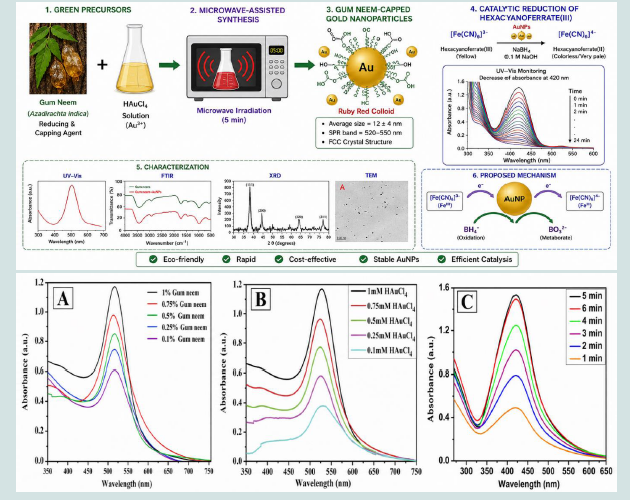 Figure 1:UV–V is absorption spectra of AuNPs synthesized using (A) different concentrations of Gum Neem, (B) different concentrations of HAuCl₄, and (C) different microwave irradiation times.
The alkaline medium (0.1 M NaOH) was used to minimize the hydrolytic decomposition of sodium borohydride and to maintain its reducing capability throughout the experiment. The progress of the reaction was monitored using a UV–Vis spectrophotometer by recording the decrease in absorbance at 420 nm, corresponding to the reduction of hexacyanoferrate(III) to hexacyanoferrate(II).
To investigate the effect of temperature on the catalytic performance, the reduction reaction was carried out over the temperature range of 30–70 °C while maintaining constant reactant concentrations. The effect of catalyst loading was also evaluated by varying the volume of the AuNP suspension from 50 to 200 μL under otherwise identical reaction conditions.
The absorption band at approximately 1600–1650 cm⁻¹, assigned to carbonyl (C=O) and carboxylate (COO⁻) groups, also exhibited a slight shift, suggesting their interaction with the AuNP surface. Similarly, the characteristic C–O and C–O–C stretching vibrations in the region of 1000–1100 cm⁻¹ showed changes in intensity and position after nanoparticle synthesis, confirming the adsorption of Gum Neem polysaccharides onto the nanoparticle surface. Minor spectral changes observed in the 1400–1450 cm⁻¹ region further support the involvement of carboxylate groups in the capping process [10-12].
Overall, these spectral changes demonstrate that the hydroxyl, carbonyl, carboxyl, and ether functional groups present in Gum Neem act as both reducing and stabilizing agents during the synthesis of AuNPs.
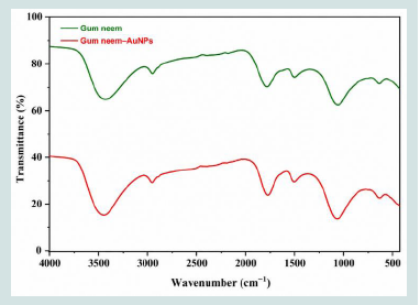 Figure 2:FTIR spectra of pure Gum Neem (GN) and Gum Neem-capped AuNPs.
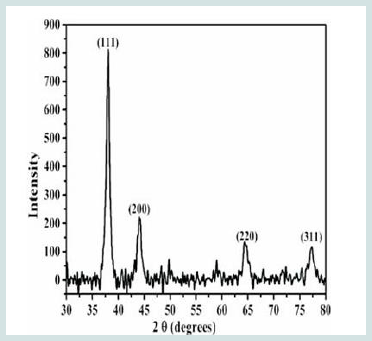 Figure 3:XRD pattern of Gum Neem-capped AuNPs synthesized using 1%
(w/v) Gum Neem and 1 mM HAuCl₄ after microwave irradiation for 5 min.
Figure 3:XRD pattern of Gum Neem-capped AuNPs synthesized using 1%
(w/v) Gum Neem and 1 mM HAuCl₄ after microwave irradiation for 5 min.
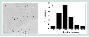 Figure 4:(A) TEM micrograph of Gum Neem-capped AuNPs and (B) the
corresponding particle size distribution histogram.
Figure 4:(A) TEM micrograph of Gum Neem-capped AuNPs and (B) the
corresponding particle size distribution histogram.
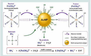 Figure 5:Proposed Mechanism of AuNPs Catalyzed Reduction of
Hexacyanoferrate (III) by NaBH₄.
Figure 5:Proposed Mechanism of AuNPs Catalyzed Reduction of
Hexacyanoferrate (III) by NaBH₄.
Both Fe(III) and Fe(II) complexes are highly stable and possess similar coordination geometries. The standard reduction potential of the Fe(III)/Fe(II) redox couple is +0.44 V, whereas borohydride oxidation occurs at −1.24 V versus the normal hydrogen electrode. Although the reaction is thermodynamically favorable, it proceeds very slowly in the absence of a catalyst because of a significant kinetic barrier. Consequently, AuNPs act as efficient electron-transfer mediators, markedly accelerating the reduction reaction.
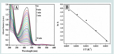 Figure 6:(A).Time-dependent UV-V is spectra of the mixture of
hexacyanoferrate (III) and sodium borohy dride upon addition of gold
nanoparticles (B) The pseudo First order kinetics of reduction₄.
Figure 6:(A).Time-dependent UV-V is spectra of the mixture of
hexacyanoferrate (III) and sodium borohy dride upon addition of gold
nanoparticles (B) The pseudo First order kinetics of reduction₄.
Research Article
Microwave-Assisted Green Fabrication of Gum Neem Capped Gold Nanoparticles for Efficient Catalytic Reduction
Banu R1* and Mohan KC2
1Department of Chemistry, Dr. BRR GDC(A) Jadcherla,
Mahabubnagar Telangana, India.
2Department of Chemistry, MALD Government Degree College Gadwal Telangana, India.
2Department of Chemistry, MALD Government Degree College Gadwal Telangana, India.
*Address for Correspondence:Ruqya Banu, Department of Chemistry, Dr. BRR GDC(A) Jadcherla, Mahabubnagar Telangana, India. EMail Id: ruqyabrr@gmail.com
Submission:20 June, 2026
Accepted:19 August, 2026
Published:22 August, 2026
Copyright: © 2026 Banu R, et al. This is an open access article
distributed under the Creative Commons Attribution License, which
permits unrestricted use, distribution, and reproduction in any medium,
provided the original work is properly cited.
Keywords: Gold Nanoparticles; Gum Neem; Green Synthesis; Catalytic
Activity; UV–Vis Spectroscopy; Biogenic Nanoparticles.
Abstract
A simple and environmentally friendly method was developed for the
green synthesis of stable gold nanoparticles (AuNPs) using Gum Neem
(GN) as both the reducing and capping agent. The synthesized AuNPs
were characterized by UV–Vis spectroscopy, Fourier-transform infrared
(FTIR) spectroscopy, X-ray diffraction (XRD), and Transmission electron
microscopy (TEM). The UV–Vis spectrum exhibited a characteristic surface
plasmon resonance (SPR) band at 520–550 nm, confirming the formation
of AuNPs. FTIR analysis revealed that the hydroxyl (–OH) functional
groups present in Gum Neem played a crucial role in the reduction
of tetrachloroauric acid to metallic gold nanoparticles. XRD analysis
confirmed the crystalline nature of the nanoparticles with a face-centered
cubic (fcc) structure, while TEM images showed uniformly dispersed, nearly
spherical nanoparticles with an average particle size of 12 ± 4 nm. The
biosynthesized AuNPs exhibited excellent catalytic activity toward the
reduction of hexacyanoferrate(III) in the presence of sodium borohydride
(NaBH₄). The reaction progress was monitored by UV–Vis spectroscopy,
and the catalytic efficiency of the AuNPs was evaluated through kinetic
studies by varying the reaction temperature and catalyst concentration.
Introduction
In recent years, noble metal nanoparticles, particularly gold
(Au), silver (Ag), and platinum (Pt), have attracted significant
research interest because of their unique physicochemical properties
at the nanoscale, which differ considerably from those of their bulk
counterparts. These properties can be precisely tailored by controlling
the size, shape, and morphology of the nanoparticles, making them
highly suitable for a broad range of technological and biomedical
applications.
Among noble metal nanomaterials, gold nanoparticles (AuNPs) have emerged as one of the most extensively investigated owing to their exceptional optical, electronic, catalytic, and biocompatible properties. Their size-dependent characteristics have enabled applications in catalysis, chemical and biological sensing, electronics, drug delivery, disease diagnosis, and other biomedical fields. In particular, AuNPs have demonstrated remarkable catalytic efficiency in electron-transfer and hydrogenation reactions, making them promising catalysts for various industrial and environmental applications [1].
Among noble metal nanomaterials, gold nanoparticles (AuNPs) have emerged as one of the most extensively investigated owing to their exceptional optical, electronic, catalytic, and biocompatible properties. Their size-dependent characteristics have enabled applications in catalysis, chemical and biological sensing, electronics, drug delivery, disease diagnosis, and other biomedical fields. In particular, AuNPs have demonstrated remarkable catalytic efficiency in electron-transfer and hydrogenation reactions, making them promising catalysts for various industrial and environmental applications [1].
Recently, biomass-derived materials have emerged as attractive
renewable resources for sustainable nanotechnology. Natural
biomass sources, including medicinal plants, agricultural wastes,
natural polymers, and other biological materials, are rich in
bioactive constituents such as polyphenols, flavonoids, alkaloids,
proteins, carbohydrates, and organic acids. These compounds act as
both reducing and stabilizing agents during the synthesis of metal
nanoparticles. Compared with conventional chemical methods,
biomass-mediated synthesis offers several advantages, including ecofriendliness,
cost-effectiveness, enhanced biocompatibility, improved
nanoparticle stability, and long-term sustainability. As a result,
biomass-derived nanomaterials have gained significant attention for
applications in catalysis, environmental remediation, sensing, energy
storage, antimicrobial and antioxidant therapies, and drug delivery
[2-6].
The catalytic performance of colloidal AuNPs is commonly
evaluated using model reduction reactions, such as the reduction
of hexacyanoferrate(III) to hexacyanoferrate(II). This reaction is
widely employed because it can be conveniently monitored by UV–
Vis spectroscopy, and the reduction proceeds at an appreciable rate
only in the presence of an efficient catalyst. Consequently, this model
reaction provides a reliable approach for evaluating the catalytic
activity and reaction kinetics of metal nanoparticles. Similar catalytic
studies have also been reported for platinum and silver nanoparticles.
Conventionally, gold nanoparticles have been synthesized
using various physical and chemical methods, including chemical
reduction, photochemical synthesis, electrochemical reduction,
thermal evaporation, and laser-assisted techniques. Although these
methods produce nanoparticles with well-controlled characteristics,
they often require expensive instrumentation, harsh reaction
conditions, and hazardous reducing or stabilizing agents such as
sodium borohydride, hydrazine, sodium citrate, and other synthetic
chemicals. The use of these toxic reagents raises concerns regarding
environmental pollution and biological safety
To overcome these limitations, green synthesis has emerged as a
sustainable and environmentally benign alternative for nanoparticle
production. Green synthetic approaches utilize naturally occurring
biomolecules from plant extracts, polysaccharides, natural gums,
microorganisms, and other renewable resources as reducing and
stabilizing agents, thereby eliminating the need for toxic chemicals.
These methods are simple, cost-effective, biocompatible, and
eco-friendly while producing stable nanoparticles with excellent
functional properties. Consequently, green synthesis has become
an attractive strategy for developing gold nanoparticles for catalytic,
biomedical, and environmental applications.
To promote sustainable nanotechnology, researchers have
increasingly focused on biological systems as environmentally friendly
alternatives for nanoparticle synthesis. Various biological resources,
including plants, bacteria, fungi, viruses, algae, agricultural waste, and
natural biopolymers, have been successfully employed as reducing
and stabilizing agents for the green synthesis of metal nanoparticles.
Among these, plant-derived natural gums have received considerable
attention because they are renewable, biodegradable, non-toxic,
and readily available. Moreover, nanoparticle synthesis using these
gums is typically carried out in aqueous media under mild reaction
conditions, eliminating the need for hazardous chemicals and making
the process economical and environmentally friendly. Several studies
have reported the successful synthesis of gold nanoparticles using
natural gums such as gum katira, kondagogu gum, and gellan gum, in
which the biomolecules act as both reducing and capping agents [7].
Gum Neem (GN), a natural exudate obtained from the stems
and branches of Azadirachta indica, is a complex polysaccharide
containing limonoids and other bioactive constituents. Owing to its
excellent biocompatibility, non-toxic nature, water solubility, and
film-forming ability, Gum Neem has been widely used as a stabilizer,
emulsifier, binder, and coating material in the food, pharmaceutical,
and cosmetic industries. The abundance of hydroxyl and other
functional groups in its molecular structure enables Gum Neem
to effectively reduce metal ions while simultaneously preventing
nanoparticle aggregation. Previous studies have demonstrated its
successful application in the green synthesis of silver and palladium
nanoparticles.
Motivated by these advantages, the present study explores the use
of Gum Neem as a dual-function reducing and stabilizing agent for the
eco-friendly synthesis of gold nanoparticles. The synthesized AuNPs
were comprehensively characterized using UV–Vis spectroscopy,
Fourier-transform infrared (FTIR) spectroscopy, X-ray diffraction
(XRD), and transmission electron microscopy (TEM). Furthermore,
the catalytic performance of the biosynthesized AuNPs was evaluated
through the electron-transfer reduction of hexacyanoferrate(III)
to hexacyanoferrate(II) in the presence of sodium borohydride.
The reaction kinetics were systematically investigated by UV–Vis
spectroscopy to assess the catalytic efficiency of the synthesized
nanoparticles.
Biosynthesis “of Gold nanoparticles employing Gum Neem extract and their potential application as” Catalytic Reduction.
Biosynthesis “of Gold nanoparticles employing Gum Neem extract and their potential application as” Catalytic Reduction.
Experimental
Materials:
Chloroauric acid trihydrate (HAuCl₄·3H₂O, 99.9%) was
obtained from Sigma-Aldrich. Gum Neem (laboratory reagent
grade, spray-dried), sodium borohydride (NaBH₄), potassium
hexacyanoferrate(III), and sodium hydroxide (NaOH) were
purchased from S. D. Fine Chemicals Ltd., Mumbai, India. All
aqueous solutions were prepared using double-distilled water, and all
chemicals were used as received without further purification.
Green Synthesis of Gum Neem-Capped Gold Nanoparticles
(GN-AuNPs)
Gold nanoparticles (AuNPs) were synthesized using Gum Neem as both the reducing and stabilizing agent. Initially, a 1% (w/v) Gum Neem stock solution was prepared by dissolving the required amount of Gum Neem powder in double-distilled water under continuous magnetic stirring for 1 h at room temperature until a homogeneous solution was obtained.
For the synthesis of AuNPs, 3 mL of the Gum Neem solution was mixed with 1 mL of 1 mM chloroauric acid (HAuCl₄) solution in a clean boiling tube. The reaction mixture was subjected to microwave irradiation for 5 min. The formation of AuNPs was confirmed by a distinct color change from pale yellow to ruby red, indicating the reduction of Au³⁺ ions to metallic gold nanoparticles.
To optimize the synthesis conditions, the concentrations of Gum Neem (0.1–1.0%, w/v) and chloroauric acid (0.1–1.0 mM) were systematically varied while maintaining all other reaction parameters constant. The synthesized nanoparticles were subsequently characterized using UV–V is spectroscopy, Fouriertransform infrared (FTIR) spectroscopy, X-ray diffraction (XRD), and transmission electron microscopy (TEM).
Gold nanoparticles (AuNPs) were synthesized using Gum Neem as both the reducing and stabilizing agent. Initially, a 1% (w/v) Gum Neem stock solution was prepared by dissolving the required amount of Gum Neem powder in double-distilled water under continuous magnetic stirring for 1 h at room temperature until a homogeneous solution was obtained.
For the synthesis of AuNPs, 3 mL of the Gum Neem solution was mixed with 1 mL of 1 mM chloroauric acid (HAuCl₄) solution in a clean boiling tube. The reaction mixture was subjected to microwave irradiation for 5 min. The formation of AuNPs was confirmed by a distinct color change from pale yellow to ruby red, indicating the reduction of Au³⁺ ions to metallic gold nanoparticles.
To optimize the synthesis conditions, the concentrations of Gum Neem (0.1–1.0%, w/v) and chloroauric acid (0.1–1.0 mM) were systematically varied while maintaining all other reaction parameters constant. The synthesized nanoparticles were subsequently characterized using UV–V is spectroscopy, Fouriertransform infrared (FTIR) spectroscopy, X-ray diffraction (XRD), and transmission electron microscopy (TEM).
Characterization of Gold Nanoparticles:
The optical properties of the synthesized AuNPs were investigated
using a UV–Vis spectrophotometer (Shimadzu UV-3600) over
the wavelength range of 200–700 nm to monitor the characteristic
surface plasmon resonance (SPR) band.FTIR spectroscopy was performed to identify the functional groups of Gum Neem involved in the reduction and stabilization of the nanoparticles. Prior to analysis, the colloidal AuNP suspension was freeze-dried, and the resulting powder was thoroughly mixed with potassium bromide (KBr) to prepare transparent pellets. A pure KBr pellet was used as the background reference. The FTIR spectra were recorded using a Shimadzu IRAffinity-1 spectrometer over the wavenumber range of 400–4000 cm⁻¹.
The crystalline structure of the synthesized AuNPs was analyzed
by X-ray diffraction using a Rigaku MiniFlex diffractometer equipped
with Cu Kα radiation.
The morphology, particle size, and particle size distribution of the synthesized AuNPs were examined using transmission electron microscopy (JEOL JEM-2000 FX-II). For TEM analysis, a drop of the colloidal AuNP suspension was placed onto a carbon-coated copper grid and allowed to dry overnight at room temperature before imaging [8].
The morphology, particle size, and particle size distribution of the synthesized AuNPs were examined using transmission electron microscopy (JEOL JEM-2000 FX-II). For TEM analysis, a drop of the colloidal AuNP suspension was placed onto a carbon-coated copper grid and allowed to dry overnight at room temperature before imaging [8].
Catalytic Reduction of Hexacyanoferrate (III):
The catalytic activity of the synthesized AuNPs was
evaluated through the electron-transfer reduction of potassium
hexacyanoferrate(III) using sodium borohydride (NaBH₄) as the
reducing agent. The reaction mixture was prepared by mixing 1.0
mL of double-distilled water, 0.5 mL of 10 mM aqueous potassium
hexacyanoferrate(III) solution, and 1.5 mL of 36 mM sodium
borohydride solution prepared in 0.1 M NaOH in a standard 3 mL
quartz cuvette. Subsequently, the required amount of the AuNP
catalyst was added to initiate the reaction.The alkaline medium (0.1 M NaOH) was used to minimize the hydrolytic decomposition of sodium borohydride and to maintain its reducing capability throughout the experiment. The progress of the reaction was monitored using a UV–Vis spectrophotometer by recording the decrease in absorbance at 420 nm, corresponding to the reduction of hexacyanoferrate(III) to hexacyanoferrate(II).
To investigate the effect of temperature on the catalytic performance, the reduction reaction was carried out over the temperature range of 30–70 °C while maintaining constant reactant concentrations. The effect of catalyst loading was also evaluated by varying the volume of the AuNP suspension from 50 to 200 μL under otherwise identical reaction conditions.
Results and Discussion
UV–Vis Spectroscopy:
UV–Vis spectroscopy is a simple, sensitive, and widely used
technique for the characterization of noble metal nanoparticles. The
formation and stabilization of gold nanoparticles (AuNPs) in aqueous
solution were monitored by UV–Vis spectroscopy. The reduction of
Au³⁺ ions to metallic Au⁰ by Gum Neem was indicated by a distinct
color change of the reaction mixture from pale yellow to ruby red.
The characteristic surface plasmon resonance (SPR) absorption band
of AuNPs was observed in the wavelength range of 520–550 nm .
[Figure 1A] shows the UV–V is spectra of AuNPs synthesized
using 1 mM chloroauric acid and varying concentrations of Gum
Neem (0.1–1.0%, w/v). As the Gum Neem concentration increased,
the intensity of the SPR band increased, indicating enhanced
formation of AuNPs.
[Figure 1B] illustrates the UV–Vis spectra obtained by varying the concentration of chloroauric acid (0.1–1.0 mM) while maintaining the Gum Neem concentration at 1% (w/v). The intensity of the SPR band increased with increasing HAuCl₄ concentration, confirming greater nanoparticle formation [9].
The effect of microwave irradiation time on AuNP synthesis was also investigated [Figure 1C]. The reaction conditions were systematically optimized to achieve maximum nanoparticle yield and stability.
[Figure 1B] illustrates the UV–Vis spectra obtained by varying the concentration of chloroauric acid (0.1–1.0 mM) while maintaining the Gum Neem concentration at 1% (w/v). The intensity of the SPR band increased with increasing HAuCl₄ concentration, confirming greater nanoparticle formation [9].
The effect of microwave irradiation time on AuNP synthesis was also investigated [Figure 1C]. The reaction conditions were systematically optimized to achieve maximum nanoparticle yield and stability.
Fourier-Transform Infrared (FTIR) Spectroscopy:
The FTIR spectra of Gum Neem-mediated gold nanoparticles
confirmed the involvement of Gum Neem biomolecules in the
reduction and stabilization of AuNPs. The broad absorption band
observed at 3200–3500 cm⁻¹, corresponding to the O–H stretching
vibration of polysaccharides, became broader and shifted slightly
after nanoparticle formation, indicating the participation of hydroxyl
groups in the reduction of Au³⁺ ions.The absorption band at approximately 1600–1650 cm⁻¹, assigned to carbonyl (C=O) and carboxylate (COO⁻) groups, also exhibited a slight shift, suggesting their interaction with the AuNP surface. Similarly, the characteristic C–O and C–O–C stretching vibrations in the region of 1000–1100 cm⁻¹ showed changes in intensity and position after nanoparticle synthesis, confirming the adsorption of Gum Neem polysaccharides onto the nanoparticle surface. Minor spectral changes observed in the 1400–1450 cm⁻¹ region further support the involvement of carboxylate groups in the capping process [10-12].
Overall, these spectral changes demonstrate that the hydroxyl, carbonyl, carboxyl, and ether functional groups present in Gum Neem act as both reducing and stabilizing agents during the synthesis of AuNPs.
X-ray Diffraction (XRD):
The crystalline structure of the synthesized AuNPs was
investigated by X-ray diffraction (XRD). As shown in Figure 3, four
distinct diffraction peaks were observed at 2θ values of 38.08°, 44.09°,
64.42°, and 77.39°, corresponding to the (111), (200), (220), and
(311) crystallographic planes, respectively. These diffraction peaks
are characteristic of the face-centered cubic (fcc) crystal structure of
metallic gold and agree well with the standard reference data (JCPDS
No. 04-0784).
The absence of additional diffraction peaks indicates the high
purity of the synthesized nanoparticles, while the sharp diffraction
peaks confirm their highly crystalline nature. The average crystallite
size, calculated using the Debye–Scherrer equation from the full width
at half maximum (FWHM) of the (111) reflection, was approximately
14 nm. This value agrees well with the particle size obtained from
TEM analysis [13-15].
Transmission Electron Microscopy (TEM):
The morphology, particle size, and size distribution of the
biosynthesized AuNPs were examined using transmission electron
microscopy (TEM). Representative TEM micrographs of AuNPs
synthesized using 1% Gum Neem and 1 mM HAuCl₄ after microwave
irradiation for 5 min are presented in [Figure 4A].
The TEM images revealed predominantly spherical nanoparticles with only a few irregularly shaped particles [16-18]. The nanoparticles were uniformly dispersed with minimal aggregation, indicating effective stabilization by Gum Neem. Analysis of individual nanoparticles showed an average particle size of 12 ± 4 nm, as illustrated in the corresponding particle size distribution histogram [Figure 4B] [19-21].
The TEM images revealed predominantly spherical nanoparticles with only a few irregularly shaped particles [16-18]. The nanoparticles were uniformly dispersed with minimal aggregation, indicating effective stabilization by Gum Neem. Analysis of individual nanoparticles showed an average particle size of 12 ± 4 nm, as illustrated in the corresponding particle size distribution histogram [Figure 4B] [19-21].
Catalytic Reduction of Hexacyanoferrate(III):
The catalytic activity of the biosynthesized AuNPs was
evaluated through the electron-transfer reduction of potassium
hexacyanoferrate(III) by sodium borohydride (NaBH₄), producing
hexacyanoferrate(II) and metaborate ions according to the following
reaction:Both Fe(III) and Fe(II) complexes are highly stable and possess similar coordination geometries. The standard reduction potential of the Fe(III)/Fe(II) redox couple is +0.44 V, whereas borohydride oxidation occurs at −1.24 V versus the normal hydrogen electrode. Although the reaction is thermodynamically favorable, it proceeds very slowly in the absence of a catalyst because of a significant kinetic barrier. Consequently, AuNPs act as efficient electron-transfer mediators, markedly accelerating the reduction reaction.
The reaction progress was monitored by UV–Vis spectroscopy.
Potassium hexacyanoferrate(III) exhibits a characteristic absorption
band at approximately 420 nm. Upon addition of AuNPs, the
absorbance at 420 nm decreased progressively with reaction time,
confirming the catalytic reduction of hexacyanoferrate(III) to
hexacyanoferrate(II) (Figure 6A). Because sodium borohydride
was present in large excess, the reaction followed pseudo-firstorder
kinetics with respect to hexacyanoferrate(III) .Throughout
the reaction, the SPR band of the AuNPs at approximately 530
nm remained essentially unchanged, indicating excellent colloidal
stability without aggregation. The absence of any significant shift or
broadening of the SPR band further demonstrates that no undesirable
interaction occurred between the reactants and the nanoparticle
catalyst.
The effect of catalyst loading was investigated by varying the
volume of AuNP suspension from 50 to 250 μL while maintaining
constant concentrations of hexacyanoferrate (III), sodium
borohydride, and temperature. The apparent rate constant increased
linearly with increasing catalyst loading, indicating that a greater
number of active surface sites enhanced electron transfer and
accelerated the reduction reaction [22-25].
The influence of temperature was also examined over the range
of 30–70 °C. As expected, the reaction rate increased with increasing
temperature because of enhanced molecular collisions and faster
electron-transfer kinetics. The activation energy (Eₐ) was determined
using the Arrhenius equation by plotting ln(k) versus 1/T (Figure
6B). From the slope (−Eₐ/R), the activation energy was calculated
to be 7.6 kcal mol⁻¹, demonstrating that the biosynthesized AuNPs
effectively lower the activation energy and facilitate rapid electrontransfer
reactions.
Conclusion
A simple, rapid, and environmentally friendly microwave-assisted
method was successfully developed for the green synthesis of gold
nanoparticles using Gum Neem as both the reducing and stabilizing
agent. The synthesized AuNPs exhibited a characteristic SPR band
at approximately 520 nm. XRD and TEM analyses confirmed the
formation of highly crystalline face-centered cubic (fcc) AuNPs
with an average particle size of 12 ± 4 nm, while FTIR spectroscopy
demonstrated that hydroxyl and carboxyl functional groups were
responsible for the reduction and stabilization of the nanoparticles.
The synthesized AuNPs exhibited excellent catalytic activity toward
the sodium borohydride-assisted reduction of hexacyanoferrate(III),
following pseudo-first-order kinetics. Increasing the catalyst loading
and reaction temperature significantly enhanced the reaction rate,
demonstrating the potential of Gum Neem-mediated AuNPs as
efficient, sustainable, and environmentally friendly nanocatalysts for
catalytic applications.
Acknowledgement
The infrastructure facilities required to carry out the experiments
were provided by Department of Chemistry Dr. B.R.R. Government
Degree College(A), Jadcherla , for which the authors is grateful.
Additionally, the authors would like to thank Prof. G. Sukanya,
Principal of “Dr. B.R.R. Government Degree College, Jadcherla,” to
provide the essential facilities and support.
References
Citation
Banu R, Mohan KC. Microwave-Assisted Green Fabrication of Gum Neem Capped Gold Nanoparticles for Efficient Catalytic Reduction. J Chem Applications 2026;6(1): 6.