
Journal of Andrology & Gynaecology
Download PDF
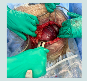 Figure 1: Intraoperative photograph of uterine torsion to the right showing
the left adnexa.
Figure 1: Intraoperative photograph of uterine torsion to the right showing
the left adnexa.
Case Report
Acute Gravid Uterine Torsion: Case Report about an Uncommon Obstetric Emergency
Slaoui A*, Lazhar H, Amail N, Zeraidi N, Lakhdar A, Baydada A and Kharbach A
Department of Gynaecology-Obstetrics & Endoscopy, Maternity
Souissi, University Hospital Center IBN SINA, University Mohammed
V, Rabat, Morocco
*Address for Correspondence:
Slaoui A, Department of Gynaecology-Obstetrics & Endoscopy, Maternity
Souissi, University Hospital Center IBN SINA, University Mohammed V,
Rabat, Morocco; E-mail: azizslaoui27@gmail.com
Submission: 10 October, 2022
Accepted: 18 November, 2022
Published: 22 November, 2022
Copyright: © 2022 Slaoui A, et al. This is an open access article
distributed under the Creative Commons Attribution License, which
permits unrestricted use, distribution, and reproduction in any medium,
provided the original work is properly cited.
Abstract
Introduction:
Uterine torsion is defined by a vertical rotation
of more than 45 degrees around its cervical-isthmic axis. It is a rare
emergency with serious complications that can be life-threatening for
the fetus and the mother. Its pathophysiology has not yet been fully
explained but this situation is generally the result of a several factors
with mainly the asymmetry of the transverse diameter of the uterus
and pelvic adhesions.
Case Presentation:
We hereby report the case of a 26-year-old
female patient, third gesture fourth pare without particular medical
history, who presented to the emergency department at 37 weeks
of amenorrhea of a twin pregnancy with severe abdominal pain
of sudden onset. Initially mistaken for retroplacental hemorrhage
or uterine rupture, it was only in the pre operatory phase of an
emergency cesarean section that the diagnosis of uterine torsion was
made. The operation was complicated by postpartum hemorrhage
due to uterine atony which was managed by prompt medical care
and a triple Tsirulnikov ligation. She was discharged home with her two
healthy newborns at D4 post-op.
Conclusion:
Uterine torsion is an uncommon and serious obstetric
complication of difficult diagnosis that can be life-threatening for the
fetus and the mother. The specificity of our case is twofold. Firstly, it
is the third case of uterine torsion in twin pregnancy reported in the
literature to date. Secondly, it is the first time to our knowledge that
the difference in length of the two round ligaments is observed as a
possible factor in the pathophysiology of this complication.
Keywords
Uterine torsion; Round ligament asymmetry; Postpartum
hemorrhage
Introduction
Uterine torsion is defined as a vertical rotation of the uterus more
than 45 degrees around its axis at the junction of the cervix and the
uterus. The first cases of uterine torsion in non-pregnant patients
with fibroids were described by Times in 1861 and Virchowen in
1863 [1]. It was not until 1876 that Labbe described the first case of
uterine torsion in a pregnant woman [2]. Since then, many cases have
been described in the literature, notably in the report of 212 cases
published by Jensen in 1992 [2]. The cases described in the published
literature generally present up to 180 degrees of torsion, but some
go up to 720 degrees [2]. This anomaly has only been observed in
three cases of twin pregnancies [3]. Clinical symptoms range from
asymptomatic to extreme abdominal pain. It is usually diagnosed late,
at the time of caesarean section. We hereby describe the third case
of uterine torsion in a twin pregnancy complicated by postpartum
hemorrhage.
Case presentation
We hereby present the case of a 26-year-old female patient,
third gesture fourth pare with two vaginal deliveries and without
particular medical history, who presented to our emergency room
with abdominopelvic pain of sudden onset at 37 weeks of amenorrhea while the course of her current twin pregnancy was without
particularities. On admission, two hours after the onset of the pain,
the patient presented with a cutaneo-mucosal heat, a conserved blood
pressure (120/60) with tachycardia at 115 bpm. She described severe
abdominal pain increased by uterine mobilization. No metrorrhagia
or loss of amniotic fluid was observed. The active fetal movements were
reduced and we noted the presence of uterine contracture. Obstetric
ultrasound revealed a still-evolving twin pregnancy with bradycardia
estimated at 80 bpm for the first twin in cephalic position and at 85
bpm for the second twin in seated position and a fundial placenta. The
suspicion of uterine rupture or retroplacental hemorrhage led to an
emergency cesarean section under general anesthesia. A non-bloody
ascites of 300 cc was observed. The uterus appeared ischemic and the
territory of the inferior segment was occupied by a vascular network
consisting mainly of ecstatic veins. We then observed perioperatively
a uterine torsion of 110 degrees to the right. A first attempt at
intra-abdominal reduction was unsuccessful and the uterus was
still presenting its left adnexa (Figure 1). A segmental Pfannenstiel
incision was made and completed with Metzenbaum chisel allowing
cephalic extraction of the first twin laterally to the abdomen and then
podalic extraction of the second twin after reduction of the uterine torsion. The fetuses weighed 2560g and 2450g respectively and had an
Apgar score of 3/5/10 and 4/6/10. The hysterorrhaphy was performed
without difficulty. There were no myomas, cysts, adhesions nor
malformations but the left round ligament (on the side contralateral
to the direction of uterine torsion) was about 5 cm longer than the
right. After easy extraction of both twins, the patient presented with
heavy bleeding despite complete artificial delivery followed by uterine
revision and rapid hysterography. Major uterine atony persisted
despite the injection of 40 IU of oxytocin. A triple Tsirulnikov
ligation was therefore performed, which made it possible to control
the postpartum hemorrhage. We completed the operative procedure
with a plication of the left round ligament. After her hemodynamic
status was restored, the patient’s postoperative course was simple.She
was discharged home with her two healthy newborns at D4 post-op.
Discussion
Uterine torsion is defined by a rotation of the uterus by more
than 45 degrees in regard to its longitudinal axis. In two thirds of
cases, it is dextrorotatoryexceeding the physiological dextrorotation
[2]. There is only one review of the literature by Jensen et al. of 212
cases [2]. In this review, fetal death in occurred in 12% of cases. Fetal
morbidity was reported, sometimes severe, but no maternal death.
Most commonly, torsion occurs during labour. Symptomatology
included metrorrhagia, cervical dystocia, uterine pain, hyperkinesia,
hypertonia, and even hemorrhagic shock [3]. Jensen et al. found that
the symptomatology is proportional to the importance of the torsion
[2]. Painful symptomatology can be concealed by peridural analgesia.
The indication for cesarean section is often based on stagnation of
dilatation or abnormalities of the fetal heart rate. Factors favoring
torsion are large uterine myomas, uterine malformations, multiple
pregnancies, or oblique or transverse fetal presentations [2]. Parity,
maternal age and gestational age do not appear to be risk factors. Our
case is characterized by a spontaneous occurrence outside labour, in
a twin pregnancy.
The etiologies that can explain this pathology are numerous
and diverse [2,4]. Among the most widely found causes are four
main categories. Firstly, the authors found situations causing an
asymmetry of the transverse diameter of the uterus, such as a
transverse presentation (22% of cases), the presence of lateralized
fibroids (21% of cases), a uterine malformation such as a bicornuate
or bifid uterus (11% of cases)and a multiple pregnancy like our case
(1% of cases). In this category we can add the important unilateral
elongation of the round ligament compared to the contralateral one
as in our case. Secondly, we have ectopic pelvic tumors, especially
ovarian (3% of cases), followed thirdly by postoperative or idiopathic
pelvic adhesions (7% of cases) and finally fourthly by morphological
abnormalities of the patient: loose abdominal wall (3% of cases), bone
abnormalities of the spine and/or pelvis (1% of cases) [2,4]. In 16%
of cases, no etiology was found [2,4]. Although all of these situations
are present in many women, torsion remains a very uncommon
obstetrical pathology. It is therefore legitimate to wonder whether a
combination of events could be at the origin of such a complication.
In 1931 Robinson and al. had already put forward this hypothesis
followed by Nesbitt et al.in 1956 [5]. The elements defined as being
the cause of this anomaly in a patient already predisposed would
be a fetal hyperactivity, false maternal movements and postural
abnormalities [2,5].
Intraoperative findings frequently include ascites and numerous
varices around the isthmic portion [3]. When the torsion exceeds 180
degrees, the diagnosis is difficult to make and the hysterotomy may
be inadvertently performed on the posterior aspect of the uterus. If
the situation is recognized prior to hysterotomy, a reduction of the
uterine torsion can be attempted in order to incise in classic territory
as we have attempted. This most often involves uterine exteriorization
[3]. If reduction is not possible, it is essential to perform the
hysterotomy away from the lateral edges of the uterus to preserve the
vascular pedicles and ureters. The choice of a vertical hysterotomy is
then preferable [3]. The risk of postpartum hemorrhage as in our case
may be increased by acute ischemia of the myometrium responsible
for atony which may lead to hemostatic hysterectomy [3]. The
literature seems to consider maternal death as exceptional but it is
certain that the occurrence of complications such as retroplacental
hemorrhage or severe postpartum hemorrhage threaten the maternal
prognosis [2,7]. For the clinician, the diagnosis of severe forms of
torsion remains difficult and the differential diagnosis is dominated
by uterine rupture or retroplacental hemorrhage.
Some authors have attempted to confirm non-acute uterine
torsion by imaging. On ultrasound, the placenta may change
position to the point of facial inversion, sometimes with ovarian
vascular anomalies [7]. Abrupt changes in placental position during
extracorporeal circulation, with maternal pain and fetal bradycardia,
have been reported as typical cases [8]. MRI can also provide another
criterion (abnormal shape of the upper vagina), but is certainly not
possible in emergency situations like our case [9]. To date, no study
has assessed the risk of recurrence after torsion and determined the
need for prophylactic uterine fixation. Fatih et al. published a case
of posterior hysterotomy for torsion on the 22nd day of pregnancy
without prior surgical intervention. Some authors recommend
plication of the round ligament [10,11]. Nevertheless, the real
usefulness of this plication remains debatable and should only be
performed when there is direct observation of obvious elongation of
a round ligament compared with the contralateral one. On the other
hand, it seems reasonable to remove large myomas before allowing a
new pregnancy.
Conclusion
Uterine torsion is an uncommon and serious obstetric
complication of difficult diagnosis that can be life-threatening for the
fetus and the mother. The specificity of our case is twofold. Firstly, it
is the third case of uterine torsion in twin pregnancy reported in the
literature to date. Secondly, it is the first time to our knowledge that
the difference in length of the two round ligaments is observed as a
possible factor in the pathophysiology of this complication.
References
Citation
Slaoui A, Lazhar H, Amail N, Zeraidi N, Lakhdar A, et al. Acute Gravid Uterine Torsion: Case Report about an Uncommon Obstetric Emergency. J Androl
Gynaecol. 2022;10(1): 3.