
Journal of Forensic Investigation
Download PDF
Research Article
*Address for Correspondence: Melissa Connor, Forensic Investigation Research Station, Colorado Mesa University, Grand Junction, Colorado 81501, USA, E-mail: mconnor@coloradomesa.edu
Citation: Couse T, Connor M. A Comparison of Maceration Techniques for Use in Forensic Skeletal Preparations. J Forensic Investigation. 2015;3(1): 6.
Copyright ©2015 Couse T, et al. This is an open access article distributed under the Creative Commons Attribution License, which permits unrestricted use,distribution, and reproduction in any medium, provided the original work is properly cited.
Journal of Forensic Investigation | ISSN: 2330-0396 | Volume: 3, Issue: 1
Submission: 06 April, 2015 | Accepted: 29 June, 2015 | Published: 03 July, 2015
A Comparison of Maceration Techniques for Use in Forensic Skeletal Preparations
Tiana Couse and Melissa Connor*
- Forensic Investigation Research Station, Colorado Mesa University, Grand Junction, Colorado 81501, USA
*Address for Correspondence: Melissa Connor, Forensic Investigation Research Station, Colorado Mesa University, Grand Junction, Colorado 81501, USA, E-mail: mconnor@coloradomesa.edu
Citation: Couse T, Connor M. A Comparison of Maceration Techniques for Use in Forensic Skeletal Preparations. J Forensic Investigation. 2015;3(1): 6.
Copyright ©2015 Couse T, et al. This is an open access article distributed under the Creative Commons Attribution License, which permits unrestricted use,distribution, and reproduction in any medium, provided the original work is properly cited.
Journal of Forensic Investigation | ISSN: 2330-0396 | Volume: 3, Issue: 1
Submission: 06 April, 2015 | Accepted: 29 June, 2015 | Published: 03 July, 2015
Abstract
Maceration removes all soft tissue from the bones. It must be done without damaging or morphing the bone. To examine the effectiveness of methods, six techniques were applied to a convenience sample of skeletal elements: simmering with two different degreasing agents (Dawn® and Greased Lightning®), power washing, insect scavenging, microwave maceration, and physical maceration. A Likert scale (modified from [1]) was used to rate the techniques. Warm water maceration with Greased Lightning® was the most effective, but warm water techniques in general rated well.Keywords
Maceration; AnthropologyIntroduction
Maceration is the act of removing all soft tissue from the bones for further examination and must be done without damaging or morphing the bone. The skeletal remains need to be clearly seen to be examined for possible causes of death due to peri- or antemortem trauma [2]. Procedures and practices of maceration were developed from many different disciplines including: museum conservation, taxidermy, and human and faunal anatomy and biology [3]. Most researchers interested in maceration are doing so for museum or curatorial purposes, and many techniques used in those venues are not suitable for a forensic setting. Some use chemicals that degrade the bone or temperatures that could compromise the cortical surface or structural integrity of the bone [3]. Generally, methods using bleach or hydrogen peroxide have been discarded due to the damage to the cortical tissue caused by the corrosive action on calcium [4].All maceration techniques aim at removing the soft tissue from the bone. Generally several steps are required: the complete removal of all soft tissue and the processes to degrease the bone and then whiten it. Initially, as much soft tissue as possible is physically removed. This includes skinning, gutting, and, usually, disarticulation in order for the bone to macerate at a more reasonable pace. For this portion of the maceration process, the preparer should have knowledge of osteology and know where the bones lie, without which bones may easily be cut or scored. In addition, the sections should be kept in labeled containers, noting the correct side. This helps ensure that information found can be equated to the correct side and area of the body, leading to more evidence from the wound.
At this point the preparer is left with a choice. Generally, techniques fall into four categories: cooking, water maceration, chemical maceration and carrion insects [4]. Dermestid beetles (a carrion insect) are a perennial favorite (for example [5]), and often used in museum settings. However, this requires a dermestid colony and significant upkeep, which is not often cost-effective for sporadic work in the forensic sciences. In addition, should the beetles escape from the colony; they can be a challenge to eradicate from a laboratory setting. So other methods are often considered.
In 2003, Fenton and others cited the “tried and true” method of submerging the specimens, adding a powdered detergent, a powdered sodium carbonate and placing the specimens over low heat, or a low simmer [4]. This process is repeated as often as needed to remove soft tissue and most of the fats, with a rinse in running water between baths. Adhering tissues are manually removed. After the final detergent bath, the remains are placed in a water and ammonia solution and cooked on low to remove grease from the bone. The ammonia expands the remaining soft tissue and makes it easier to remove.
Reflecting the rising interest in preserving DNA and other cellular components of the bone, Steadman and her colleagues experimented with different maceration methods and tested whether DNA remained after the bone was cleaned [3]. They discuss the use of bleach, hydrogen peroxide, ethylenediaminetetraacetic acid (EDTA)/papain, and room temperature water and detergent/sodium carbonate, all followed by the use of degreasing agents. They conclude that treatments performed at high temperatures (90° or above) for short durations generally yielded the best post-maceration DNA results.
Simonsen and others tried commercial enzymatic products (three different proteases and a lipase) and found that the enzymes significantly speeded up the process over the traditional warm water maceration [6]. King and Birch were concerned with the preservation of cut marks on the bone post-maceration and discuss manual maceration, chemical and enzymatic solutions used with a warm water bath, invertebrate methods (insects), and cooking using microwaves, simmering and boiling [1]. They found no single maceration technique applied to all situations.
A very gentle technique, suitable for fetal and infant material is described by Love and Sanchez [7]. They use an incubator to aid in warm water maceration as the machine was able to heat the bath temperature evenly without creating hotspots, as happens when hot plates are used. The detergent used was a 1:2 ratio of foremost 1553- ES Super Kleen® (Delta Foremost Chemical Corporation, Memphis TN) and water.
After all of the soft tissue is removed, the bone should further be degreased. However, bleach degrades bone and can remove the outer layers [8]. Ammonia then can be used to help whiten the bones or simply dry them out more for a prolonged use.
When working with any tools or chemicals it is important to keep in mind the safety of the preparer. Exposure to only minute amounts of toxic chemicals can cause damage to cells [9]. All safety precautions should be followed (Table 1). This includes wearing appropriate clothing for the lab, face wear, and using a fume hood when heating any chemicals, even household detergents.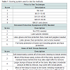 The purpose of the present project was to examine maceration techniques to use at Colorado Mesa University’s Forensic Investigation Research Station. The first author is both a student at CMU and employed at a taxidermist where she regularly macerates skeletal remains for clients. In that venue, she was also using a power washer to remove soft tissue and that technique has not previously been reported in the literature.
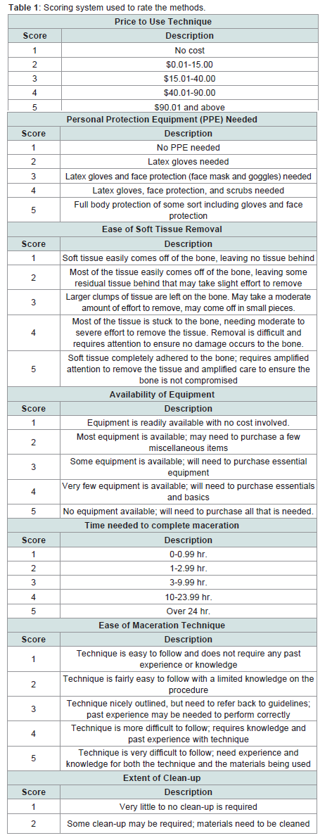
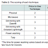
Table 1: Scoring system used to rate the methods.
The techniques chosen for testing were based on the availability of materials, how long each technique would take, and techniques currently used in a forensic context. This comparison allows the testing to be within real-world parameters plausible during a forensic case.
The materials used in the study were a convenience sample, meaning that they were chosen without regard to probability sampling, but because of their accessibility to the researcher. The sample consisted five mountain lion skulls and two full bear paws, and two sets of human remains. While wild feline and bear bones differ from human bone in shape, they still consist of collagen and hydroxyapatite crystal and the maceration techniques should have the same or similar effects.
The feline skulls were attained from a local taxidermist who needed the skulls of the cats cleaned for his clients. Care was taken to ensure that the bone was not compromised during the skinning process and that minimal to no soft tissue was removed. Each skull was similar in size, and the amount of soft tissue on each was observed. The bear paws were donated to Colorado Mesa University’s Forensic Investigation Research Station (FIRS) for student study and comparison with human hands. No animals were killed for this study. The paws were dismembered above the carpals and the skin was removed. Each paw had the same amount of soft tissue remaining on it. The human remains were donated to FIRS for taphonomic research.
Physical maceration
Physical maceration only requires latex gloves, forceps, a scalpel and a scouring pad. Once the latex gloves were on, the scalpel was used to remove large chunks of soft tissue and the forceps were used to pull off smaller pieces. A scouring pad was used over the bone to remove remaining pieces of tissue. Water was used to clean smaller pieces still adhered to the bone.
Power washing
Power washing could be considered a form a physical maceration, applying water dispensed under pressure to remove the tissue. A Hotsy® power washer, a rubber protection suit, rubber boots, goggles, latex gloves, and a face mask, was used. The technique also requires a safe area for the detritus from the spraying. In this case a concrete pad surrounded by two foot high walls was used. Once the operator donned the PPE, the bone was placed in the safe area and the power washer was connected to an electrical outlet and a water tap (via hose). When connected, the skull was sprayed until the tissue was removed and then set out to dry.
Warm water maceration techniques
Dawn® dish soap and Greased Lightning®:The materials needed include a fume hood, crock pot, Dawn® dish soap, water, forceps, and latex gloves. The crock pot was filled with tap water and one tablespoon of Dawn® dish soap. The mixture was left to simmer at a constant 100 °C with the fleshed bone for 24 hours in two 12 hour shifts. Gloves and forceps were then used to remove the bone from the water and a double straining system (colander over a screen over the drain) was used to strain the dirty water. Running water was poured on the soft tissue to cool it off. The same process was used for maceration with Greased Lightning®, which the authors obtained from a local hardware store.
Microwave maceration: Microwave maceration requires a 1,000 watt microwave, a bowl, water, and latex gloves. The bone is submerged in the bowl of water and placed into the microwave. The microwave was run on high for five minutes, being monitored every ten seconds for soft tissue changes. Running water was used to rinse off the bones.
Outside maceration with and without tarp
Outside maceration with and without tarp: Two sets of human remains donated to FIRS were placed in the outdoor facility. Both were placed outside on March 6, 2015 within 15 m of each other. One was placed under a tarp and the other placed outside without a tarp. Both were nude and supine. The donation placed outside without a tarp was female and not autopsied; the donation placed under the tarp was male and had been autopsied; the autopsy cut had not been sewn up. Most remains at FIRS quickly desiccate beyond the point attractive to blowfly larva and the concept was that the tarp would keep the remains moist and allow the larva to complete more of the removal of the soft tissue.
A scoring system was adapted from King and Birch and used to compare the techniques (Table 1) [1]. Seven criteria were used in the scoring system: price, personal protection equipment (PPE) needed, the ease of the removal of soft tissue, the availability of equipment, the time needed for full maceration, the ease of the technique itself, and the amount of cleanup needed after the technique was used. These criteria were chosen based on the practical applications of each technique in a laboratory. The scoring was from 1 (best) to 5 (worse), so a lower score indicates a better technique (Table 2,Table 3).
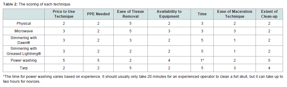
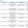
Table 2: The scoring of each technique.
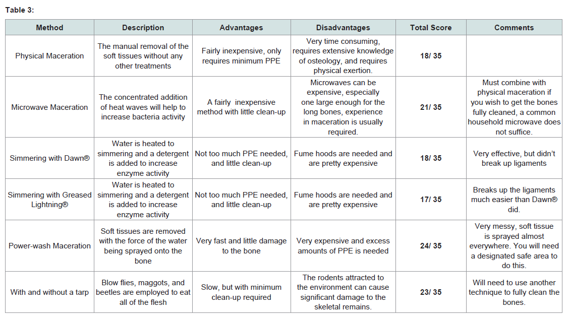
Table 3:
Results
Based on the total scores of each method, warm water maceration with Greased Lightning® scored the best. It is easy to use, fairly inexpensive, and comes with little clean-up after the maceration is completed (Table 2,Table 3). However, the technique is time consuming and the use of a fume hood is required because some materials can get very odoriferous when they are simmering. These results agrees with other research showing procedures that involve heating to 90 °C or higher clean bones faster [10]. This can be observed with both simmering techniques, the microwave technique, and also the outdoor maceration because the warmer the day, the more insect activity was observed.Physical maceration
Physical maceration is by far the least expensive technique and requires the least amount of PPE. However, there are significant drawbacks. The technician performing this technique should be sophisticated in osteology. Otherwise, it is far too easy to unintentionally cut or mark the bone when trying to remove the tissue. It is also very time consuming and a fair amount of physical activity is needed. The flesh is completely adhered to the bone and the macerator uses just a scalpel and scouring pad to remove the flesh. This technique worked best when paired with another technique to soften the flesh or partially remove it.
Power washing
This technique is very quick. With minimal experience with a power washing machine, an operator can remove the soft tissues from the bone with ease. However, the machine is very expensive, high levels of PPE are needed, and a dedicated area to clean the bone is required as pieces of tissue tend to be sprayed everywhere. The force of the power washer can knock teeth out of a skull. The bone is easily cleaned, but the high pressure of the water can also damage it. Weaknesses in the bone due to the very defects for which a forensic scientist is looking, may be the areas damaged during power washing. Due to this damage, this technique is unusable for a forensic setting, but may work very nicely for other less delicate settings, including taxidermy.
Warm water maceration
Simmering with Greased Lightning®: This technique had the best results in the ease of tissue removal. The flesh fell off the bear paws easily without much physical removal of tissue needed afterword. However, it did take a couple days, generated a considerable amount of odor, and required multiple baths to fully macerate the paws and the adhering ligaments still required scraping to remove. Greased Lightning®® softened the ligaments as well as the muscle tissue, making the removal of those tougher tissues much easier than other warm water maceration techniques.
Simmering with Dawn® dish soap: Simmering with Dawn® also resulted in easy removal of the soft tissues. Dawn® worked well to remove the softer tissue, but did not soften the ligaments as well as Greased Lightning®. Both warm water maceration techniques require a fume hood as they are fairly odoriferous and the chemicals in these detergents should not be inhaled.
Microwave maceration: This technique was the least useful of the warm water maceration techniques. First, the size of the bone that could be used was severely constricted by the size and shape of the microwave. Then, it took an hour of microwaving to soften the flesh, with no discernable soft tissue removal. This technique was time consuming and ultimately ended in having to use physical maceration to fully clean the skull.
Outdoor maceration with and without tarp
The decomposition of the bodies were scored on the scale developed by Megysi and others where 3 is a fresh body, six through 16 are stages of early decomposition with bloating, marbling, and color changes to a black or brown tone, 17 to 24 are stages of advanced decomposition, and 24 to 35 are stages of skeletonization (Table 4) [11]. Initially, the body under the tarp decomposed more quickly, going through the stages of early decomposition faster than the body in the open. This is probably due to the fact that the bodies were laid out in March when the weather was cool and the black plastic covering raised the temperature under the tarp increasing micro bacterial activity. Both sets of remains showed significant maggot activity, but on neither were significant areas of skeleton exposed by the insects.
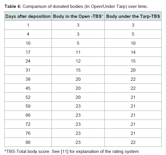
Table 4: Comparison of donated bodies (In Open/Under Tarp) over time.
The body in the open progressed slightly faster in late decomposition and at Day 80, the body under the tarp showed more moist decomposition than the body in the open. On Day 80, the body under the tarp did have more visible beetle activity than the one in the open, but did not show significantly more exposed skeleton. During late decomposition, the body under the tarp also became host to a mouse nest in the chest (Figure 1). While rodent activity is seen commonly on the donations at FIRS, this set of remains under a tarp is the only one to date where the rodents built a nest in the body. The tarp clearly made conditions more hospitable for the rodents, as well as the insects.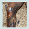 Clearly, waiting more than 80 days to macerate the remains for a forensic case is rarely acceptable. Nor is the damage to the skeletal elements that will likely be caused by the rodents, as rodents commonly leave gnaw marks on skeletal elements.
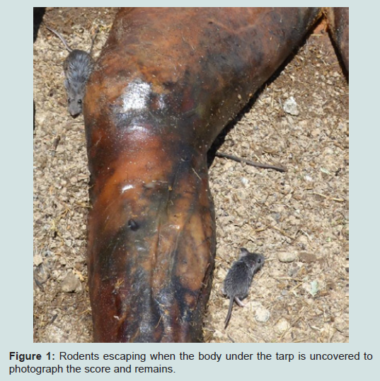
Figure 1: Rodents escaping when the body under the tarp is uncovered to photograph the score and remains.
Discussion
Warm water maceration with Greased Lightning®, followed by an ammonia bath proved the simplest and most cost-effective method used in this suite of techniques. The power washing, given a suitable outdoor work space, is quite quick and effective, but has the down sides of working solely on sturdy bone, needing significant PPE for the researcher, and when working with human materials - a dedicated space that can literally be sprayed with bio-hazardous materials.Laying out a body in a dedicated space and letting nature take its course is attractive. In western Colorado and other areas where humidity is low, however, nature tends to produce mummies more often than skeletons not useful when the goal is maceration. Even excluding large scavengers, the damage that can be done to the skeletal elements through avian and rodent scavengers is significant. This is useful for taphonomic studies, less useful for forensic casework.
This study, despite the added techniques and different goals, came close to the conclusions expressed by Steadman and others on the most effective maceration technique [3]. King and Birch favored the microwave method for preservation of bone and cut mark [1]. The present study found that the microwave technique is slow and limited in the size of the skeletal element. It would be difficult to heat, for example, the femur of an adult male in a normal sized microwave. The King and Birch study used ribs from adult pigs cut into sections of three adjacent ribs, each five cm long. It would be inappropriate to cut forensic case skeletal elements into microwave-sized pieces, and thus this utility of this approach is limited.
King and Birch did make an excellent point in concluding that no one maceration technique meets all criteria [1]. For the most part, those who conduct macerations wish the bone to remain intact, the process to add no additional marks on the bone, and the process not to destroy the integrity of any marks that were on the bone before the maceration. Warm water maceration seems to be the best current technique to do that. The suggestion by Steadman and others that shorter term immersion in water of higher temperature will yield a better chance of preserving DNA is also a criteria to keep in mind [3].
Conclusion
Simmering with Greased Lightning® scored the best of the techniques used in this study. Using this technique, soft tissues were easily removed without causing damage or deforming the bone. The most effective method would ultimately depend on the case and the resources available to the lab handling said case. This experiment does provide a clearly outlined explanation of each technique and the results of its use. Based on this, it could help future technicians to decide which technique(s) would be best applied to their case.Acknowledgements
The authors thank the donors of the human material and their families. Gregory Brumfield is thanked for allowing use of his facilities and for supplying many of the materials needed to complete this study. Also thanks to Dr. John Williams at Western Carolina University who first told me about using Greased Lightning®.References
- King C, Birch W (2015) Assessment of maceration techniques used to remove soft tissue from bone in cut mark analysis. J Forensic Sci 60: 124-135.
- Mairs S, Swift B, Rutty GN (2004) Detergent: An alternative approach to traditional bone cleaning methods for forensic practice. Am J Forensic Med Pathol 25: 276-284.
- Steadman DW, DiAntonio LL, Wilson JJ, Sheridan KE, Tammariello SP (2006) The effects of chemical and heat maceration techniques on the recovery of nuclear and mitochondrial DNA from bone. J Forensic Sci 51: 11-17.
- Fenton TW, Birkby WH, Cornelison J (2003) A fast and safe non-bleaching method for forensic skeletal preparation. J Forensic Sci 48: 274-276.
- Hefti E, Trechsel U, Rufenacht H, Fleisch H (1980) Use of dermestid beetles for cleaning bones. Calcif Tissue Int 31: 45-47.
- Simonsen KP, Rasmussen AR, Mathisen P, Petersen H, Borup F (2011) A fast preparation of skeletal materials using enzyme maceration. J Forensic Sci 56: 480-484.
- Love JC, Sanchez LA (2009) Recognition of skeletal fractures in infants: an autopsy technique. J Forensic Sci 54: 1443-1446.
- Rennick SL, Fenton TW, Foran DR (2005) The effects of skeletal preparation techniques on DNA from human and non-human bone. J Forensic Sci 50: 1016-1019.
- Galloway A, Snodgrass JJ (1998) Biological and chemical hazards of forensic skeletal analysis. J Forensic Sci 43: 940-948.
- Lee EJ, Luedtke JG, Allison JL, Arber CE, Merriwether DA, et al. (2010) The effects of different maceration techniques on nuclear DNA amplification using human bone. J Forensic Sci 55: 1032-1038.
- Megyesi MS, Nawrocki SP, Haskell NH (2005) Using accumulated degree-days to estimate the postmortem interval from decomposed human remains. J Forensic Sci 50: 618-626.