
Journal of Oral Biology
Download PDF
Research Article
*Address for Correspondence: Dr. Iryna Branets, DDS., Department of Cariology and Comprehensive Care, NYU College of Dentistry, New York, NY, Tel: (718) 8910994, (917)5898533, (718) 934 0050; E-mail: iob200@nyu.edu
Citation: Branets I, Stabulas J, Dauer LT, Quinn B, Dauer Z, et al. Pediatric Bitewing Exposure to Organs of the Head and Neck Through the Use of Juvenile Anthropomorphic Phantoms. J Oral Bio. 2014;1(1): 5.
Copyright © 2014 Branets et al. This is an open access article distributed under the Creative Commons Attribution License, which permits unrestricted use, distribution, and reproduction in any medium, provided the original work is properly cited.
Journal of Oral Biology | ISSN: 2377-987X | Volume: 1, Issue: 1
Submission: 02 September 2014 | Accepted: 17 October 2014 | Published: 20 October 2014
Pediatric Bitewing Exposure to Organs of the Head and Neck Through the Use of Juvenile Anthropomorphic Phantoms
Branets I*, Stabulas J, Dauer LT, Quinn B, Dauer Z, Miodownick D, Hershkowitz DH, Colosi DC and Goren AD
- New York University College of Dentistry, Stony Brook University School of Dental Medicine, Memorial Sloane-Kettering Cancer Center, USA
*Address for Correspondence: Dr. Iryna Branets, DDS., Department of Cariology and Comprehensive Care, NYU College of Dentistry, New York, NY, Tel: (718) 8910994, (917)5898533, (718) 934 0050; E-mail: iob200@nyu.edu
Citation: Branets I, Stabulas J, Dauer LT, Quinn B, Dauer Z, et al. Pediatric Bitewing Exposure to Organs of the Head and Neck Through the Use of Juvenile Anthropomorphic Phantoms. J Oral Bio. 2014;1(1): 5.
Copyright © 2014 Branets et al. This is an open access article distributed under the Creative Commons Attribution License, which permits unrestricted use, distribution, and reproduction in any medium, provided the original work is properly cited.
Journal of Oral Biology | ISSN: 2377-987X | Volume: 1, Issue: 1
Submission: 02 September 2014 | Accepted: 17 October 2014 | Published: 20 October 2014
Abstract
Background: A literature review revealed that there is a lack of valid data on bitewing exposure dosimetry to the head and neck organs of pediatric patients. Here we are determining the actual dose to two anthropomorphic juvenile (5 yr old and 10 yr old) phantoms.Objective: To yield dosimetric measurements on the X-ray exposures to the organs of the head and neck from bitewing radiographs taken of two juvenile CIRS phantoms using round and rectangular collimation for both film and digital radiography.
Methods and Materials: Two anthropomorphic CIRS juvenile phantoms (5 yr old and 10 yr old) were exposed for bitewing radiographs using a Gendex 765 x-ray machine at the manufacturers pre-set film and digital pediatric settings for both rectangular and round collimation. Optically Stimulated Luminescence (OSL) dosimeters were placed in 21 head and neck pre-manufactured slots in each phantom. All exposures were repeated 15 times and divided by 15 to evaluate the average dose per view. Average organ dose calculated in micro Gray was based on 4 bitewing views. Organ fractions irradiated were determined from ICRP-89. Organ equivalent doses and overall effective doses (micro Sieverts) were based on 4 bitewing views and the ICRP-103 tissue weighting factors.
Results: Radiation exposures using rectangular and round collimators were about the same for both phantoms. The effective dose in micro Sieverts for the 5 year old ranged from a low of 1.8 (digital rectangular) to a high of 3.1 (film-round). The 10 year old ranged from a low of 1.5 micro Sieverts (digital-rectangular) to a high of 2.8 (film rectangular) and 2.7 (film-round). Thyroid and other organ doses were low, with the highest doses seen in the glands, extrathoracic airway, and the oral mucosa.
Conclusions: This was the first study utilizing juvenile CIRS phantoms in conjunction with OSL dot dosimetry to provide organ dose data from pediatric bitewing radiographs. Digital imaging and rectangular collimation along with pediatric machine settings must be used so that the ALARA concept in relation to children’s head and neck exposure may be maximized.
Introduction
Claus [1] and co-authors recently reported an association between bitewing radiographs and meningiomas that relied on patient’s recall on events that occurred in the past. A patient’s recall of past dental radiographic history is not an accurate means of assessing data accurately and could result in erroneous conclusions. We must admit that there could be a slim possibility linking dental x-rays to meningiomas, since prior to ALARA there was no rectangular collimation, faster speed films, or digital radiography. Unfortunately, the study lacked valid statistical data on the actual patient exposure dosimetry. A literature review also revealed that there was no valid data on bite wing dosimetry to the head and neck organs of pediatric patients.Intraoral dental radiographs are a necessary diagnostic tool in the treatment of oral diseases, such as caries, oral pathologies and periodontal disease [2]. Posterior bitewing (BW) x-rays are recognized as an important tool for the diagnosis of dental developmental stages from childhood through adulthood [3]. As they image the coronal portion of the tooth and the alveolar crestal bone in both arches so that the interproximal areas might be visualized for carious lesions [4,6].
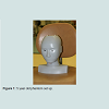
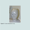
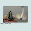
Most of the studies on radiation doses from BW x-rays in the literature have been performed using phantoms of adult males and thermoluminescent dosimetry (TLD). The current study addresses juveniles (5 and 10 year olds) (Figure 1) based on four bite wing views are performed. The study utilized optically stimulated (OSLs) which have been shown to perform better at low level dental radiation doses than TLDs [7,8]. OSLs were positioned throughout the different layers of the head and neck to measure the doses for BW exposures using a combination of digital (Figure 2), F-speed film, round collimation and rectangular collimation. Average tissue-absorbed dose, tissue equivalent dose and overall effective dose were calculated following International Commission on Radiological Protection (ICRP) recommendations (Figure 3).
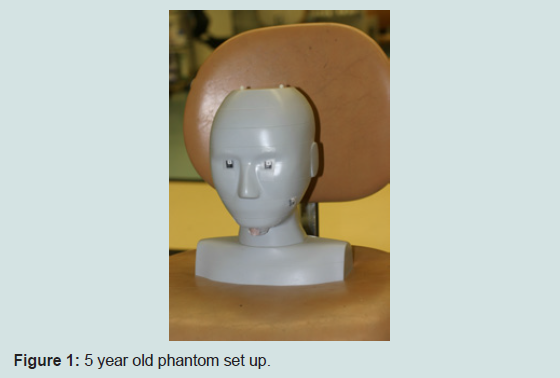
Figure 1: 5 year old phantom set up.
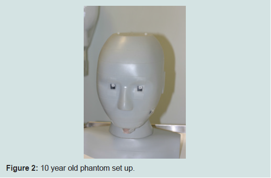
Figure 2: 10 year old phantom set up.
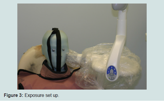
Figure 3: Exposure set up.
Objective
To yield dosimetric measurements on the X-ray exposures to the organs of the head and neck from bitewing radiographs taken of two juvenile (5 year old and 10 year old) Computerized Imaging Reference System (CIRS) phantoms using round and rectangular collimation for both film and digital radiography.Materials and Methods
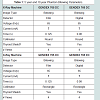
Two anthropormorphic CIRS juvenile phantoms (5 years old and 10 years old) were exposed for bitewing radiographs using a Gendex 765 x-ray machine at the manufacturers pre-set film and digital pediatric settings for both rectangular and round collimation (Table 1). The phantoms were designed specifically to ICRP anatomical references. Optically Stimulated Luminescence (OSL) dosimeters (4 mm diameter), thin (<0.5 mm) were placed in 21 head and neck pre-manufactured slots in each phantom. The OSL dosimeters were made of plastic discs infused with aluminum oxide doped with carbon (NanoDot OSLs). All exposures were repeated fifteen times to provide more precise measure of radiation to the dosimeters since it was anticipated that the individual absorbed doses per image would be below the minimal detection limit of the dosimeters, and the results were divided by 15 to evaluate the average dose per view. The OSLs were read using laser stimulation in a reader (MicroStar™ Inlight Reader) calibrated to the X-ray energies utilized. For both phantoms, F-speed film and digital radiographic settings were utilized when using round or rectangular collimation. A 6 cm diameter position indicating device with a length of 30 cm was used for round collimation techniques. For rectangular collimation, a diaphragm collimator insert with a 3.6x4.6 cm2 opening with a length of 30 cm was used. Lead thyroid shields with 0.5 mm lead equivalent were employed for all exposures. Average organ dose was calculated in micro Grays based on four bitewing views. Organ fractions irradiated were determined from ICRP-89 [9]. Organ equivalent doses and overall effective doses (micro Sieverts) were based on four bitewing views and the ICRP-103 tissue weighting factors.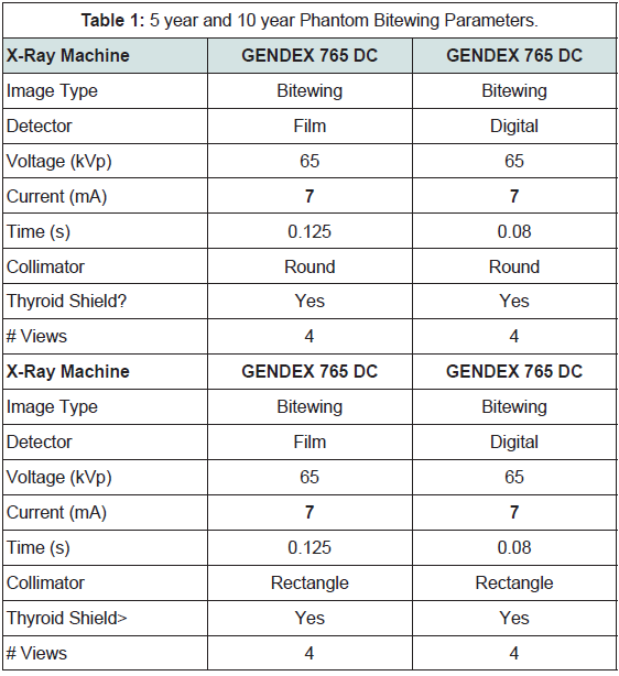
Table 1: 5 year and 10 year Phantom Bitewing Parameters.
Results
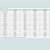
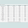
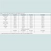
For the 5 year old the dose to the brain with round collimation/film was 6.93E-01 micro Grays, with round collimation/digital the organ dose was 5.73E-01 micro Grays. When the exposure for film was used for rectangular collimation the organ dose was reduced to 1.24E+00 and to 6.73E-01 for the digital/rectangular exposure (Table 2). With the 10 year old the organ doses to the brain were 3.05E-01 micro Grays for round/film, 3.18E-01 micro Grays for round/ digital, 6.42E-01 micro Grays for rectangular/film, and 2, 77E-03 micro Grays for rectangular/digital (Table 3). Overall the radiation exposures using rectangular and round collimators were about the same for both phantoms. The effective dose in micro sieverts for the 5 year old ranged from a low of 1.8 (digital/rectangular) to a high of 3’1 (round/film). The 10 year old ranged from a low of 1.5 micro Sieverts (digital/rectangular) to a high of 2.8 (film/rectangular) and 2.7 (film/ round) (Table 4). Doses to the thyroid and other organs were low. The highest doses were seen in the glands, extrathoracic airway, and the oral mucosa.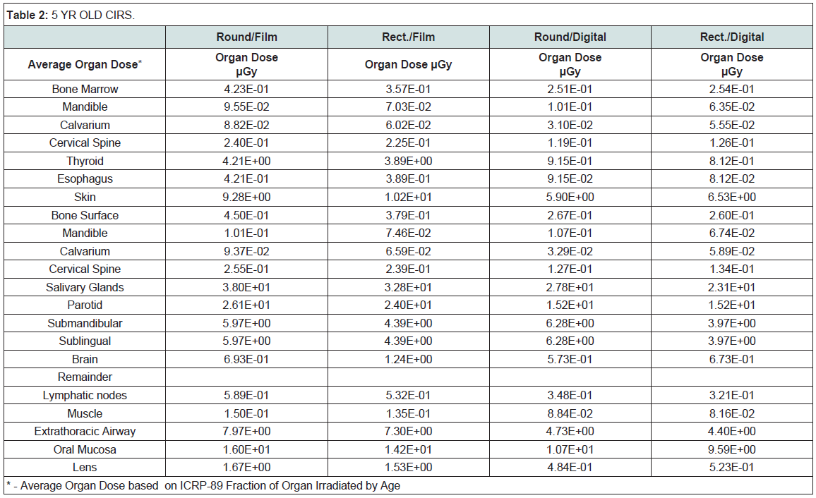
Table 2: 5 year Old CIRS.
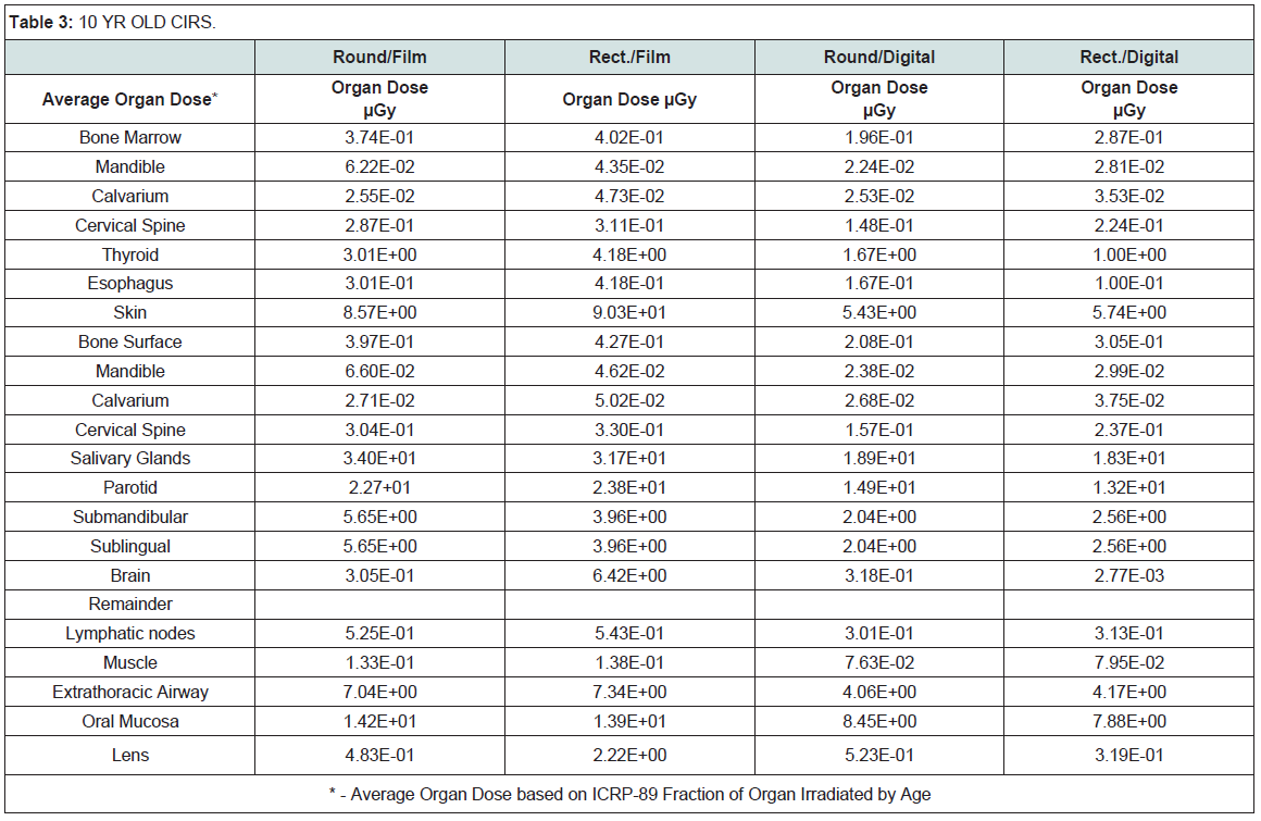
Table 3: 10 year Oid CIRS.
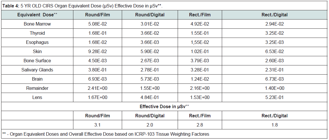
Table 4: 5 year Old CIRS Organ Equivalent Dose (μSv) Effective Dose in μSv**.
Discussion
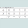
This is the first study to employ anthropomorphic juvenile phantoms consistent with ICRP recommendations and is the first to utilize OSL technology for accurately measuring absorbed dose in tissue for BW examinations. The principles of justification and radiation safety and protection must be adhered to. This study provides information that is necessary to ensure that when BW radiography is justified, the procedure is also optimized so that the radiation doses are as low as reasonably achievable (ALARA) to obtain the necessary diagnostic information. Rectangular collimation reduced the effective dose in children by approximately 4-10%. The use of time setting for digital imaging versus film imaging (F-speed) reduced the effective dose by almost 40%. When both digital imaging and rectangular collimation were employed the effective dose was reduced approximately 43%. Therefore the effective doses can be reduced by a factor of 2 for children. This study confirms that when ALARA principles are used, (digital detectors, rectangular collimation, leaded thyroid shield and child-size exposure times) patient exposure is reduced Table 5.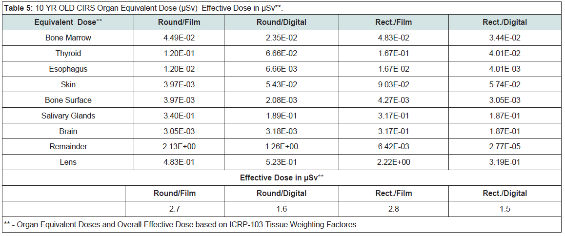
Table 5: 10 year Old CIRS Organ Equivalent Dose (μSv) Effective Dose in μSv**.
Conclusions
The results of this study reinforces past recommendations of the American Dental Association regarding simple and effective ways to reduce patient exposure. An object of a good dental radiographic quality assurance program is to ensure that doses are kept ALARA while maintaining that diagnostic information is obtained. For children the Image Gently philosophy must be adhered to and our findings confirmed that digital imaging in conjunction with rectangular collimation must be used so that the ALARA concept in relation to children’s head and neck exposure may be maximized.References
- Claus EB, Calvocoressi L, Bondy ML, Schildkraut JM, Wiemels JL, et al. (2012) Dental x-rays and risk of meningioma. Cancer 118: 4530-4537.
- ADA (2012) Dental radiographic examinations: recommendations for patient selection and limiting radiation exposure. American Dental Association, Council on Scientific Affairs. U.S. Department of Health and Human Services, Public Health Service, Food and Drug Administration.
- USFDA (2004) The selection of patients for dental radiographic examinations. U.S. Food and Drug Administration.
- Wenzel A (2004) Bitewing and digital bitewing radiography for detection of caries lesions. J. Dent. Res 83 Spec No C: C72-5.
- Brooks, S.L (2009) Guidelines for Prescribing Dental Radiographs. Mosby.
- Button TM, Moore WC, Goren AD (1999) Causes of excessive bitewing exposure: results of a survey regarding radiographic equipment in New York. Oral Surg Oral Med Oral Pathol Oral Radiol Endod 87: 513-517.
- Valiyaparambil JV, Mallya SM (2011) Characterization of an optically stimulated dosimeter for dentomaxillofacial dosimetry. Oral Surg Oral Med Oral Pathol Oral Radiol Endod 112: 793-797.
- Branets I, Colosi DC, Branets L, Quinn B, Dauer L, T Fernandez, et al. (2014). Optically stimulated luminescence and thermoluminescence dosimetry: an overall perspective. Research 1: 720.
- Stabin M, Emmons MA, Segars WP, Fernald M, Brill AB (2008) ICRP-89 based adult and pediatric phantom series. J Nucl Med 49: 14P.