
Journal of Forensic Investigation
Download PDF
Research Article
*Address for Correspondence: Daniel Vanek, Forensic DNA Service, Janovskeho 18, 17000 Prague 7, Czech Republic, Tel: +420 603 979 915; Fax: +420 233 931 123; E-mail: daniel.vanek@DNA.com.cz
Citation: Zatkalikova L, Bazovsky R, Turanska M, Vanek D. DNA Analysis of Human Skeletal Remains in Slovakia: Laboratory Set-Up, Success Rates and Compliance of the Procedures with the ISO Guidelines. J Forensic Investigation. 2014;2(1): 11.
Copyright © 2014 Zatkalikova L, et al. This is an open access article distributed under the Creative Commons Attribution License, which permits unrestricted use,distribution, and reproduction in any medium, provided the original work is properly cited.
Journal of Forensic Investigation | ISSN: 2330-0396 | Volume: 2, Issue: 1
Submission: 14 January 2014 | Accepted: 10 March 2014 | Published: 14 March 2014
DNA Analysis of Human Skeletal Remains in Slovakia: Laboratory Set-Up, Success Rates and Compliance of the Procedures with the ISO Guidelines
Livia Zatkalikova1, Richard Bazovsky1, Martina Turanska1 and Daniel Vanek2,3*
- 1Institute of Forensic Science of Slovak Police Forces, Bratislava, Slovak Republic
- 2Forensic DNA Service, Prague, Czech Republic
- 3Charles University, 2nd Faculty of Medicine, Institute of Legal Medicine, Czech Republic
*Address for Correspondence: Daniel Vanek, Forensic DNA Service, Janovskeho 18, 17000 Prague 7, Czech Republic, Tel: +420 603 979 915; Fax: +420 233 931 123; E-mail: daniel.vanek@DNA.com.cz
Citation: Zatkalikova L, Bazovsky R, Turanska M, Vanek D. DNA Analysis of Human Skeletal Remains in Slovakia: Laboratory Set-Up, Success Rates and Compliance of the Procedures with the ISO Guidelines. J Forensic Investigation. 2014;2(1): 11.
Copyright © 2014 Zatkalikova L, et al. This is an open access article distributed under the Creative Commons Attribution License, which permits unrestricted use,distribution, and reproduction in any medium, provided the original work is properly cited.
Journal of Forensic Investigation | ISSN: 2330-0396 | Volume: 2, Issue: 1
Submission: 14 January 2014 | Accepted: 10 March 2014 | Published: 14 March 2014
Abstract
Aim: To evaluate the performance of a police laboratory that specializes in the DNA identification of skeletal remains in terms of success rates and compliance of procedures with international quality assurance standards and recommendations; to describe the correlations between sample type, post-mortem interval and level of decomposition of ostheological samples and autosomal STR typing in a dataset of 219 individuals (323 samples) in Slovakia; and to provide readers with comprehensive guidelines for the set-up and operation of a specialized bone identification DNA laboratory. This evaluation should serve as a reference for all planned specialized laboratories for the DNA identification of bone samples.Method: Skeletal remains were recovered from different locations throughout Slovakia, and the post-mortem intervals ranged from several days to 84 years. The preservation of these samples varied depending on the storage conditions prior to recovery. Therefore, the silica-based DNA extraction method that was successfully used in the identification of victims from mass graves in former Yugoslavia was used here as the method of choice.
Results: The comparison of results from specific years indicated constant improvement in the overall success rate, from 71% in 2005/2006 to a maximum of 100% in 2009. There were several factors that influenced the success rate: a robust extraction procedure, continuous improvement of methods, bone sample selection, the introduction of DNA quantification and strict counter-cross contamination procedures.
Conclusion: The results of this study clearly demonstrate that the proper application of scientifically sound procedures can lead to the efficient identification of unknown skeletal remains, thus solving serious criminal cases. The success of DNA typing was related to the type and the post-mortem interval of the skeletal sample; thus, the proper education of officers submitting case samples can result in an increase in the overall success rate as well as speed up the process of identification.
Introduction
The escalation in crime rate that occurred after the Velvet Revolution in Czechoslovakia (1989) caused an increase in the demand for forensic DNA laboratories, which are powerful resources that can help the police identify unknown bodies using molecular methods. The plan of the Police of the Slovak Republic was to set up a specialized DNA laboratory dedicated to the identification of skeletal remains of victims of brutal crimes and mass fatality incidents, and this plan was supported by the positive results of the identification efforts in the former Yugoslavia [1] and the World Trade Center [2]. The Slovak bone laboratory began its operation based on the original protocol published by Davoren et al. [3], and the results published herein summarize the cases that were solved between November 2005 and December 2011.Material and Methods
Laboratory set-upThe Slovak Bone DNA Identification Laboratory is located at the Institute of Forensic Science, Slovenska Lupca. This police DNA laboratory performs all types of forensic DNA typing and possesses a special room for DNA extraction from bone specimens involved in criminal cases. The laboratory space dedicated to DNA extraction from bone samples is separated from the remaining laboratory area by a double door. The bone extraction laboratory is under positive pressure, with a filtered exhaust and decontamination system located outside the building. The 15-m2 room is equipped with a sink, strong UV lamps (254 nm) and laboratory tables. The list of instruments required for successful DNA extraction from bone samples is shown in DNA extraction method section. This set-up allows for the processing of up to 10 samples per day.
Counter cross-contamination procedures
Procedures for the maintenance of bio-clean status were adopted from a standard operating procedure for DNA extraction from bone samples [3].
The bone extraction laboratory is cleaned at the end of the day. The laboratory area is regularly and systematically monitored to check for DNA contamination and, when detected, is removed through appropriate decontamination techniques using a commercial bleach solution. The presence of background DNA contamination is assessed using extraction negative controls.
Only bone samples submitted for forensic examination are processed in the dedicated laboratory. No positive extraction controls or other high-molecular-weight DNA samples are allowed to enter this laboratory space.
Extraction negative controls are used, and all suspicious qRTPCR or STR analysis results are recorded. When possible, all samples are processed in duplicate (extraction A and extraction B).
Trained specialists are allowed to enter the dedicated laboratory area, and disposable lab coats or coveralls, gloves, breathing masks, and head coverings must be worn the entire time they are in that area.
Instruments, tools and boxes of consumables that are to be placed in the dedicated laboratory are cleaned with a 5% bleach solution, water and isopropyl alcohol before they are brought into the room.
Only molecular biology grade solutions and unopened original extraction buffers are used in this laboratory.
Wherever possible, only plastic consumables that are certified to be human DNA-free, PCR-clean or, at the very least, bio-pure or bioclean are used. Only DNA LoBind 1.5- and 2.0-mL tubes are used (Eppendorf, Germany). All plastic ware, including tips, tubes and concentrators, are UV-irradiated for 20 minutes in a CL-1000 UV crosslinker (UVP, USA) before use. Only filtered tips are used.Sample typesSamples submitted for forensic DNA analysis vary in age and quality. The post-mortem interval (PMI) of the skeletal remains included in this study ranged from several days (burnt miners) to 84 years (skeletal remains in a crypt). A summary of the PMIs is shown in (Figure1). The majority of the cases were connected to killings (52%), homicides (20%), public menaces (13%) and manslaughter (7%).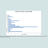
Sample Preparation for DNA Extraction
Dirt was removed manually from the surface of all samples,except for teeth, and bone samples that were sanded using a Dremel tool (Dremel, USA). The sample surface was then sanded down to remove a layer 1-2 mm thick. Samples were subsequently cut into smaller pieces, and sample fragments were placed in a 50-mL tube and cleaned using bleach-water-ethanol washing steps. Once cleaned, the sample fragments were placed into a clean 50-mL tube and were completely dried in a vacuum concentrator (Eppendorf, Germany) that was placed in a laminar hood. Three different grinding procedures were used to transform the fragments into a fine powder. An IKA mill (IKA Werke, Sweden), a Warring mill (Warring, USA) or a 2-ton press with stainless cups (Amplicon, Czech Republic) was used, depending on the sample type. The press was useful for tooth samples, whereas the other two mills were used for bone samples.
DNA Extraction Method
DNA extraction from skeletal remains of different ages is based on the methodology published by the International Commission on Missing Persons [3], with some modifications. The changes to the original protocol were introduced to reflect the variable status of the submitted samples; for example, the “teeth protocol” uses lower buffer volumes.
DNA extraction protocol was performed using the following instruments: Multipurpose centrifuge 5810 (Eppendorf), Rotor A-4- 81 (Eppendorf), Adapter 5x50 mL (Eppendorf), Adapter 12x15 mL (Eppendorf), Flex bucket – MTP (Eppendorf), Adapter 24x1.5 mL (Eppendorf), Thermomixer comfort (Eppendorf), Exchangeable thermoblock 4x50 ml (Eppendorf), Exchangeable thermoblock 24x1.5 mL (Eppendorf), Vacuum concentrator (Eppendorf), Pipettors (Eppendorf), Biohazard laminar flow hood (Ekokrok,Slovak Republic), Scales 0.1 g (Kern, Germany), Dremel tool + accessories (Dremel), Sieve Mill (IKA, Sweden), Blender with 75 mL cup (Waring, USA), 2-ton press and stainless cups (Amplicon, Czech Republic), CL-1000 UV crosslinker (UVP, USA).
The following set of chemicals was used for DNA extraction from bone samples according to the protocol described below: ATL buffer (Qiagen, Germany), AL buffer (Qiagen), AW1 buffer (Qiagen), AW2 buffer (Qiagen), AE buffer (Qiagen), proteinase K (Sigma, USA), DTT (dithiothreitol) (Sigma), PA buffer (Amplicon), 96% ethanol (Sigma), Molecular biology grade water (Sigma), Bleach (sodium hypochloride (Bochemie, Czech Republic), and DNAstable (Biomatrica, USA).
DNA extraction protocol consists of 41 steps: (the tooth protocol buffer volumes are shown in parentheses/italics): pre-heat ATL buffer (55°C) in the thermomixer; put 1.5 g of bone powder in a 50-mL tube; add 15 mL (5.0 mL) of ATL buffer; add 1 μL (300 μL) of proteinase K (20 mg/ml); add 250 μl (80 μL) of 1 M DTT; incubate 18-24 h at 55°C in a thermomixer (200 rpm); pre-heat AL buffer (55°C); add 16.5 mL (5.4 mL) of AL buffer; incubate at 70°C for at least 1 h, spin at 3,000 rpm for 5 min; label new 50-mL tube with sample number; add 16.5 ml (5.4 mL) of absolute ethanol; transfer the supernatant to the tube with the ethanol (for DNA extraction protocol with a modified step including the addition of PA buffer (Amplicon) add 5 μL of PA buffer (Amplicon); mix well; apply at least one third (the whole volume) of the sample to the maxi spin column; spin at 3,000 rpm for 3 min; discard flow-through; repeat previous 3 steps two more times; discard flow-through; apply 10 mL (5.0 mL) AW1 buffer to the spin column; spin at 3,000 rpm for 15 min; transfer the spin column to a new 50-mL tube labeled with the sample number; pre-heat AE buffer to 70°C; apply 3 mL of AE buffer to the column; incubate for 5 min at 70°C; spin at 3,000 rpm for 2 min; apply 3 mL of AE buffer to the column; Leave 5 min at room temperature; spin at 3,000 rpm for 10 min; transfer the sample to an Amicon Ultra-15 Centrifugal Filter Unit (100 kDa) (Millipore, USA); spin at 3,000 rpm for 3 min; wash the sample with 6 mL of water; spin at 3,000 rpm for 3 min; wash the sample with 6 mL of water; Spin at 3,000 rpm for 2 min;transfer sample to a DNA-free 1.5-mL tube; concentrate the sample to a final volume of 50 μL using a vacuum concentrator; store at -20°C for a maximum of 2 months; For sample storage longer than 2 months, store in a -70°C deep freezer; for sample storage longer than 2 months, use DNAstable (Biomatrica).
DNA Quantitation, Amplification and Capillary Electrophoresis
Extracted DNA was quantified using the Quantifiler™ Human DNA Quantification Kit (Applied Biosystems, USA) on a Real Time- PCR System ABI PRISM® 7900 HT (Applied Biosystems, USA) according to manufacturers’ instructions.
STR amplification was performed using an Eppendorf MCEP Gradient S thermocycler (Eppendorf). The following PCR amplification kits were used in combination with AmpliTaq Gold® DNA polymerase (Applied Biosystems): PowerPlex® 16 System(Promega Corporation, USA), PowerPlex® 16 HS (Promega Corporation), PowerPlex® Y system (Promega Corporation), AmpFlSTR® Identifiler™ (Applied Biosystems), AmpFlSTR® Yfiler®(Applied Biosystems), and AmpFlSTR® Yfiler® (Applied Biosystems). All PCR amplification kits were used according to manufacturer’ recommendations.
Amplified DNA was separated using ABI 310 and 3130 genetic analyzers (Applied Biosystems).
 A total of 35 bone samples were extracted in duplicate using a modified protocol that included the addition of PA buffer (Amplicon) to extraction B. PA buffer (Amplicon) contains carrier RNA fragments that are 50-100 bases in length. Table 2 shows the results for 37 sample pairs with and without the addition of PA buffer (Amplicon). In total, 31 samples had the same amplification success rates, whereas 5 samples amplified better after the addition of PA buffer (Amplicon), and 1 sample failed to amplify after the addition of PA buffer (Amplicon). The primary function of PA buffer (Amplicon) is to increase the binding of small DNA fragments to the silica matrix.
Counter cross-contamination procedures
Procedures for the maintenance of bio-clean status were adopted from a standard operating procedure for DNA extraction from bone samples [3].
The bone extraction laboratory is cleaned at the end of the day. The laboratory area is regularly and systematically monitored to check for DNA contamination and, when detected, is removed through appropriate decontamination techniques using a commercial bleach solution. The presence of background DNA contamination is assessed using extraction negative controls.
Only bone samples submitted for forensic examination are processed in the dedicated laboratory. No positive extraction controls or other high-molecular-weight DNA samples are allowed to enter this laboratory space.
Extraction negative controls are used, and all suspicious qRTPCR or STR analysis results are recorded. When possible, all samples are processed in duplicate (extraction A and extraction B).
Trained specialists are allowed to enter the dedicated laboratory area, and disposable lab coats or coveralls, gloves, breathing masks, and head coverings must be worn the entire time they are in that area.
Instruments, tools and boxes of consumables that are to be placed in the dedicated laboratory are cleaned with a 5% bleach solution, water and isopropyl alcohol before they are brought into the room.
Only molecular biology grade solutions and unopened original extraction buffers are used in this laboratory.
Wherever possible, only plastic consumables that are certified to be human DNA-free, PCR-clean or, at the very least, bio-pure or bioclean are used. Only DNA LoBind 1.5- and 2.0-mL tubes are used (Eppendorf, Germany). All plastic ware, including tips, tubes and concentrators, are UV-irradiated for 20 minutes in a CL-1000 UV crosslinker (UVP, USA) before use. Only filtered tips are used.Sample typesSamples submitted for forensic DNA analysis vary in age and quality. The post-mortem interval (PMI) of the skeletal remains included in this study ranged from several days (burnt miners) to 84 years (skeletal remains in a crypt). A summary of the PMIs is shown in (Figure1). The majority of the cases were connected to killings (52%), homicides (20%), public menaces (13%) and manslaughter (7%).
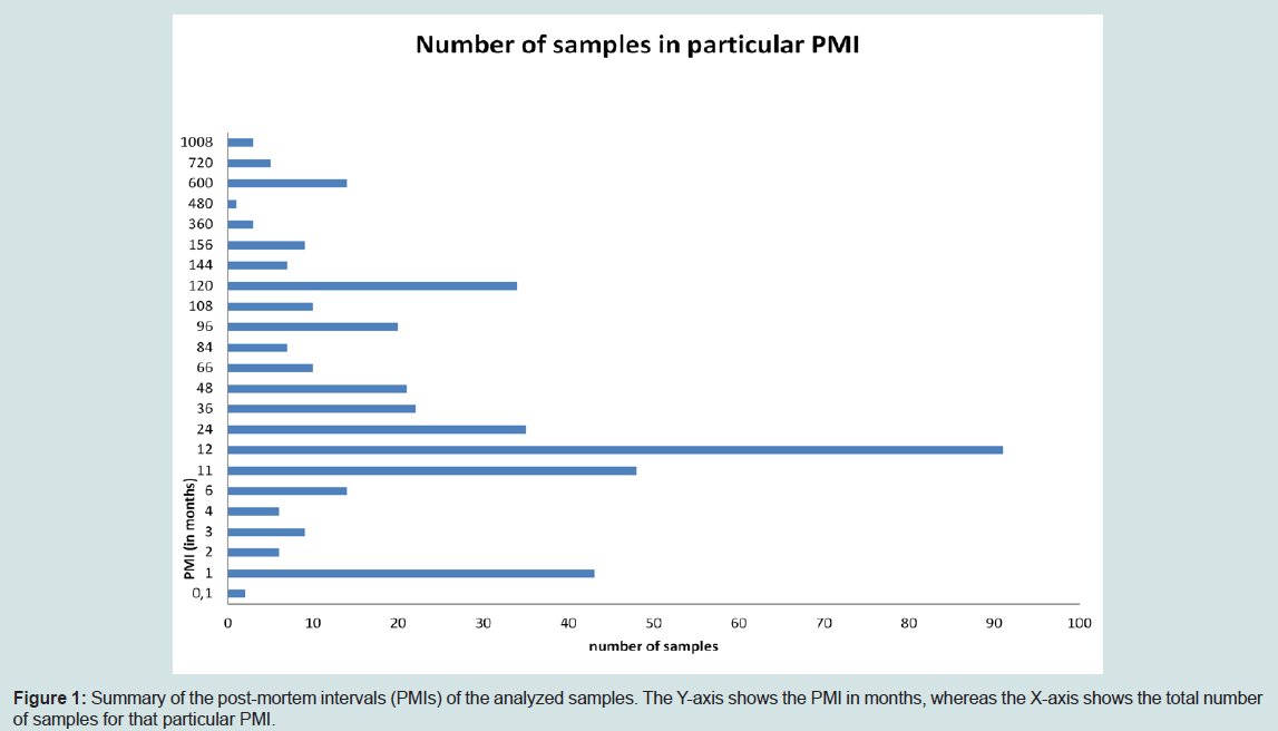
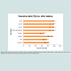
Figure 1: Summary of the post-mortem intervals (PMIs) of the analyzed samples. The Y-axis shows the PMI in months, whereas the X-axis shows the total number of samples for that particular PMI.
Sample Preparation for DNA Extraction
Dirt was removed manually from the surface of all samples,except for teeth, and bone samples that were sanded using a Dremel tool (Dremel, USA). The sample surface was then sanded down to remove a layer 1-2 mm thick. Samples were subsequently cut into smaller pieces, and sample fragments were placed in a 50-mL tube and cleaned using bleach-water-ethanol washing steps. Once cleaned, the sample fragments were placed into a clean 50-mL tube and were completely dried in a vacuum concentrator (Eppendorf, Germany) that was placed in a laminar hood. Three different grinding procedures were used to transform the fragments into a fine powder. An IKA mill (IKA Werke, Sweden), a Warring mill (Warring, USA) or a 2-ton press with stainless cups (Amplicon, Czech Republic) was used, depending on the sample type. The press was useful for tooth samples, whereas the other two mills were used for bone samples.
DNA Extraction Method
DNA extraction from skeletal remains of different ages is based on the methodology published by the International Commission on Missing Persons [3], with some modifications. The changes to the original protocol were introduced to reflect the variable status of the submitted samples; for example, the “teeth protocol” uses lower buffer volumes.
DNA extraction protocol was performed using the following instruments: Multipurpose centrifuge 5810 (Eppendorf), Rotor A-4- 81 (Eppendorf), Adapter 5x50 mL (Eppendorf), Adapter 12x15 mL (Eppendorf), Flex bucket – MTP (Eppendorf), Adapter 24x1.5 mL (Eppendorf), Thermomixer comfort (Eppendorf), Exchangeable thermoblock 4x50 ml (Eppendorf), Exchangeable thermoblock 24x1.5 mL (Eppendorf), Vacuum concentrator (Eppendorf), Pipettors (Eppendorf), Biohazard laminar flow hood (Ekokrok,Slovak Republic), Scales 0.1 g (Kern, Germany), Dremel tool + accessories (Dremel), Sieve Mill (IKA, Sweden), Blender with 75 mL cup (Waring, USA), 2-ton press and stainless cups (Amplicon, Czech Republic), CL-1000 UV crosslinker (UVP, USA).
The following set of chemicals was used for DNA extraction from bone samples according to the protocol described below: ATL buffer (Qiagen, Germany), AL buffer (Qiagen), AW1 buffer (Qiagen), AW2 buffer (Qiagen), AE buffer (Qiagen), proteinase K (Sigma, USA), DTT (dithiothreitol) (Sigma), PA buffer (Amplicon), 96% ethanol (Sigma), Molecular biology grade water (Sigma), Bleach (sodium hypochloride (Bochemie, Czech Republic), and DNAstable (Biomatrica, USA).
DNA extraction protocol consists of 41 steps: (the tooth protocol buffer volumes are shown in parentheses/italics): pre-heat ATL buffer (55°C) in the thermomixer; put 1.5 g of bone powder in a 50-mL tube; add 15 mL (5.0 mL) of ATL buffer; add 1 μL (300 μL) of proteinase K (20 mg/ml); add 250 μl (80 μL) of 1 M DTT; incubate 18-24 h at 55°C in a thermomixer (200 rpm); pre-heat AL buffer (55°C); add 16.5 mL (5.4 mL) of AL buffer; incubate at 70°C for at least 1 h, spin at 3,000 rpm for 5 min; label new 50-mL tube with sample number; add 16.5 ml (5.4 mL) of absolute ethanol; transfer the supernatant to the tube with the ethanol (for DNA extraction protocol with a modified step including the addition of PA buffer (Amplicon) add 5 μL of PA buffer (Amplicon); mix well; apply at least one third (the whole volume) of the sample to the maxi spin column; spin at 3,000 rpm for 3 min; discard flow-through; repeat previous 3 steps two more times; discard flow-through; apply 10 mL (5.0 mL) AW1 buffer to the spin column; spin at 3,000 rpm for 15 min; transfer the spin column to a new 50-mL tube labeled with the sample number; pre-heat AE buffer to 70°C; apply 3 mL of AE buffer to the column; incubate for 5 min at 70°C; spin at 3,000 rpm for 2 min; apply 3 mL of AE buffer to the column; Leave 5 min at room temperature; spin at 3,000 rpm for 10 min; transfer the sample to an Amicon Ultra-15 Centrifugal Filter Unit (100 kDa) (Millipore, USA); spin at 3,000 rpm for 3 min; wash the sample with 6 mL of water; spin at 3,000 rpm for 3 min; wash the sample with 6 mL of water; Spin at 3,000 rpm for 2 min;transfer sample to a DNA-free 1.5-mL tube; concentrate the sample to a final volume of 50 μL using a vacuum concentrator; store at -20°C for a maximum of 2 months; For sample storage longer than 2 months, store in a -70°C deep freezer; for sample storage longer than 2 months, use DNAstable (Biomatrica).
DNA Quantitation, Amplification and Capillary Electrophoresis
Extracted DNA was quantified using the Quantifiler™ Human DNA Quantification Kit (Applied Biosystems, USA) on a Real Time- PCR System ABI PRISM® 7900 HT (Applied Biosystems, USA) according to manufacturers’ instructions.
STR amplification was performed using an Eppendorf MCEP Gradient S thermocycler (Eppendorf). The following PCR amplification kits were used in combination with AmpliTaq Gold® DNA polymerase (Applied Biosystems): PowerPlex® 16 System(Promega Corporation, USA), PowerPlex® 16 HS (Promega Corporation), PowerPlex® Y system (Promega Corporation), AmpFlSTR® Identifiler™ (Applied Biosystems), AmpFlSTR® Yfiler®(Applied Biosystems), and AmpFlSTR® Yfiler® (Applied Biosystems). All PCR amplification kits were used according to manufacturer’ recommendations.
Amplified DNA was separated using ABI 310 and 3130 genetic analyzers (Applied Biosystems).
Quality Assurance Issues
The experts working in the bone extraction laboratory were trained and certified for DNA identification of bone samples at the Forensic DNA Service, Prague, Czech Republic. The Slovak Police bone DNA identification laboratory is accredited according to ISO 17025:2005 and regularly undergoes GEDNAP trials as well as proficiency testing of bone samples with unknown DNA profiles.Results
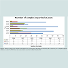
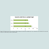
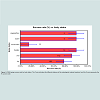
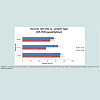
A total of 323 samples from 219 individuals were processed from November 2005 to December 2011. The skeletal remains were recovered from different locations of the Slovak Republic, and the post-mortem interval ranged from several days to 84 years (see Figure1). Other variables in addition to PMI that influenced the robustness of DNA identification were site status (see Figure 2) and body status (wet, dry, burnt, cooked, mummified, etc.), as shown in Table1. The quality of some of samples was also influenced by the maceration technique used by the Institute of the Legal Medicine prior to sample submission. Figure 3 summarizes the different crimes and mass fatality incidents that resulted in the submission of skeletal remains. The state of preservation of these samples varied depending on storage conditions prior to recovery. Thus, the silica-based DNA extraction method that was successfully used in the identification of victims from mass graves was used as the method of choice. Figure 4 shows the number of different sample types submitted to the laboratory for processing during the period from November 2005 to December 2011. The majority of the samples were teeth (62.52%) and femur samples (20.4%), and the remaining samples comprised several other sample types. Figure 5 presents the numbers of sample types (teeth, femur, other) divided by year to simplify presentation. The number of tooth samples submitted increased each year. Figure 6 shows the DNA typing success rate according to sample type, whereas Figure 7 shows the dependence of the DNA typing success rate on the PMI. Figure 8 shows the relationship between success rates and burial sites, while Figure 9 shows the relationship between the DNA typing success rate and body status. Figure 10 compares success rates (%) with sample types after the introduction of the qRT-PCR assay.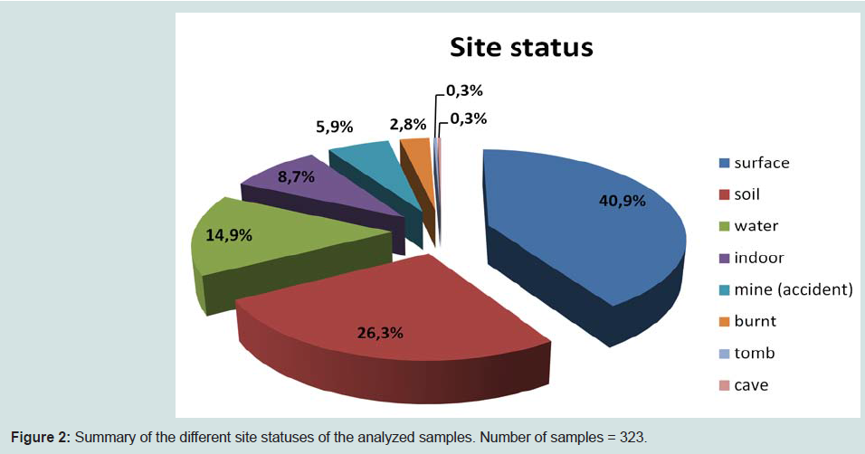
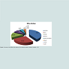
Figure 2: Summary of the different site statuses of the analyzed samples. Number of samples = 323.
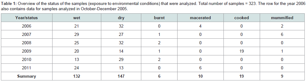
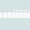
Table 1: Overview of the status of the samples (exposure to environmental conditions) that were analyzed. Total number of samples = 323. The row for the year 2006 also contains data for samples analyzed in October-December 2005.
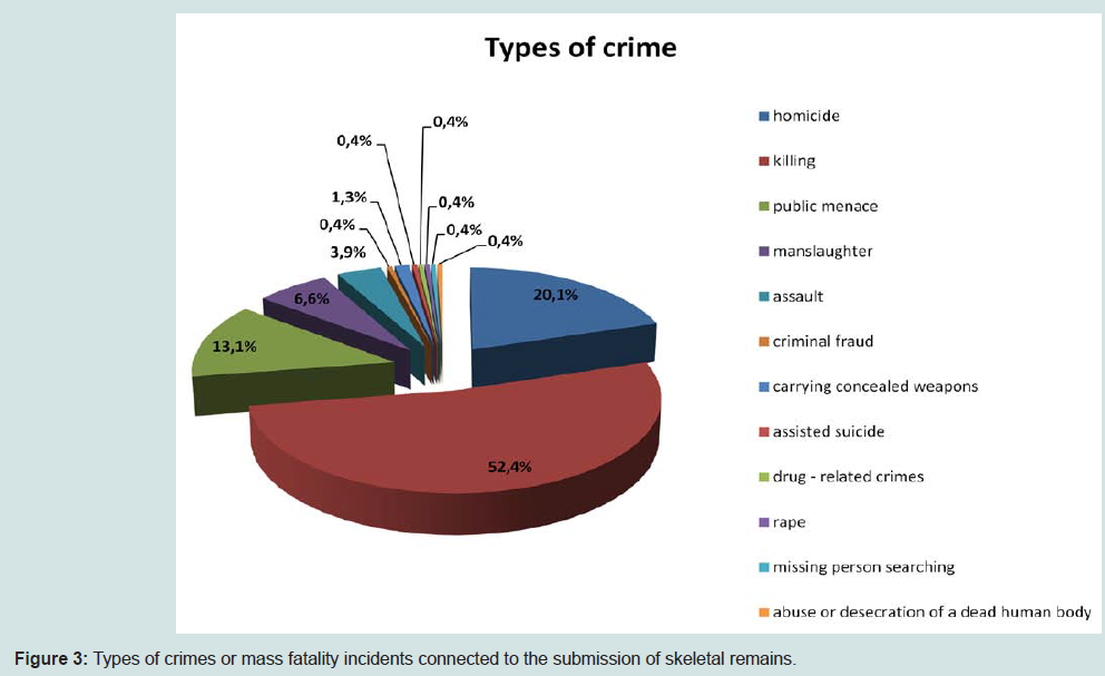
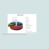
Figure 3: Types of crimes or mass fatality incidents connected to the submission of skeletal remains.
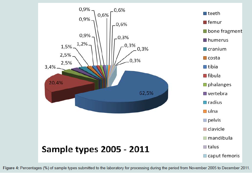
Figure 4: Percentages (%) of sample types submitted to the laboratory for processing during the period from November 2005 to December 2011.
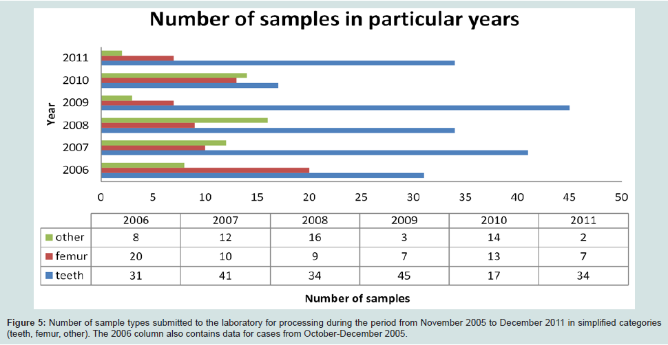
Figure 5: Number of sample types submitted to the laboratory for processing during the period from November 2005 to December 2011 in simplified categories (teeth, femur, other). The 2006 column also contains data for cases from October-December 2005.z
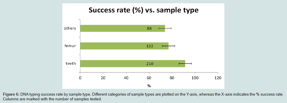
Figure 6: DNA typing success rate by sample type. Different categories of sample types are plotted on the Y-axis, whereas the X-axis indicates the % success rate. Columns are marked with the number of samples tested.
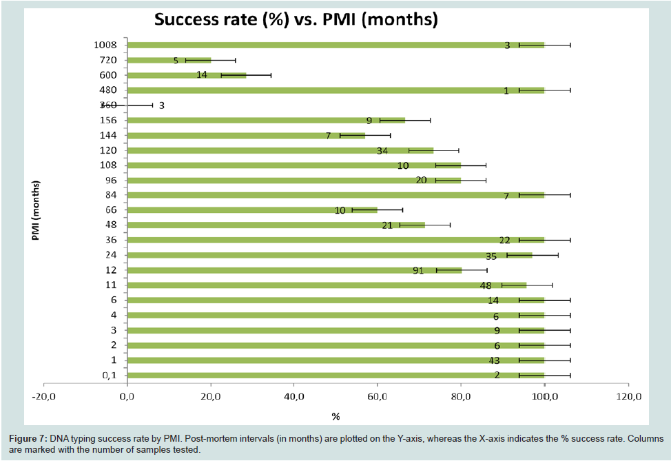
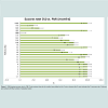
Figure 7: DNA typing success rate by PMI. Post-mortem intervals (in months) are plotted on the Y-axis, whereas the X-axis indicates the % success rate. Columns are marked with the number of samples tested.
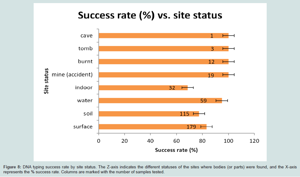
Figure 8: DNA typing success rate by site status. The Z-axis indicates the different statuses of the sites where bodies (or parts) were found, and the X-axis represents the % success rate. Columns are marked with the number of samples tested.
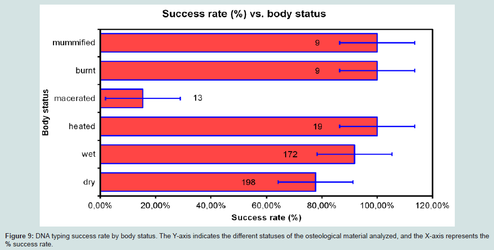
Figure 9: DNA typing success rate by body status. The Y-axis indicates the different statuses of the osteological material analyzed, and the X-axis represents the% success rate.
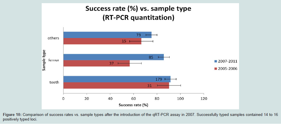
Figure 10: Comparison of success rates vs. sample types after the introduction of the qRT-PCR assay in 2007. Successfully typed samples contained 14 to 16 positively typed loci.
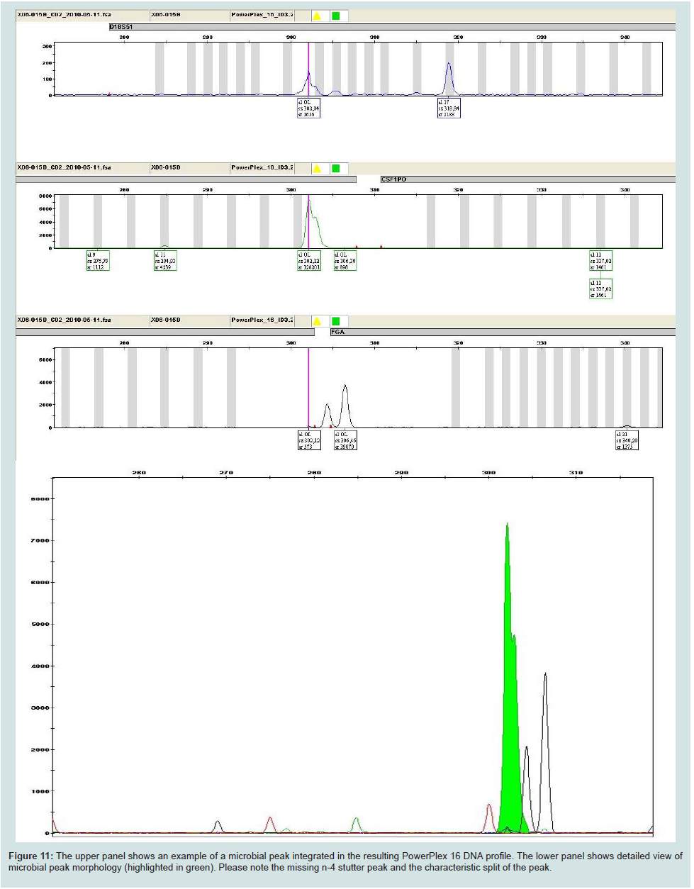
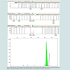
Figure 11: The upper panel shows an example of a microbial peak integrated in the resulting PowerPlex 16 DNA profile. The lower panel shows detailed view of microbial peak morphology (highlighted in green). Please note the missing n-4 stutter peak and the characteristic split of the peak.
Table 2: Effect of PA buffer (Amplicon, Czech Republic) addition on the amplification success rate. The last column lists the post-mortem interval (PMI) in months. Forensic samples with small amounts of DNA are prone to contamination with foreign DNA. One sample out of the 238 samples processed in this study (teeth, indoor, dry, PMI 120 months) showed signs of contamination. Subsequent laboratory checks excludedthe laboratory staff as sources of contamination; however, the spin columns used, which are not certified as human DNA-free, were a possible source of contamination.
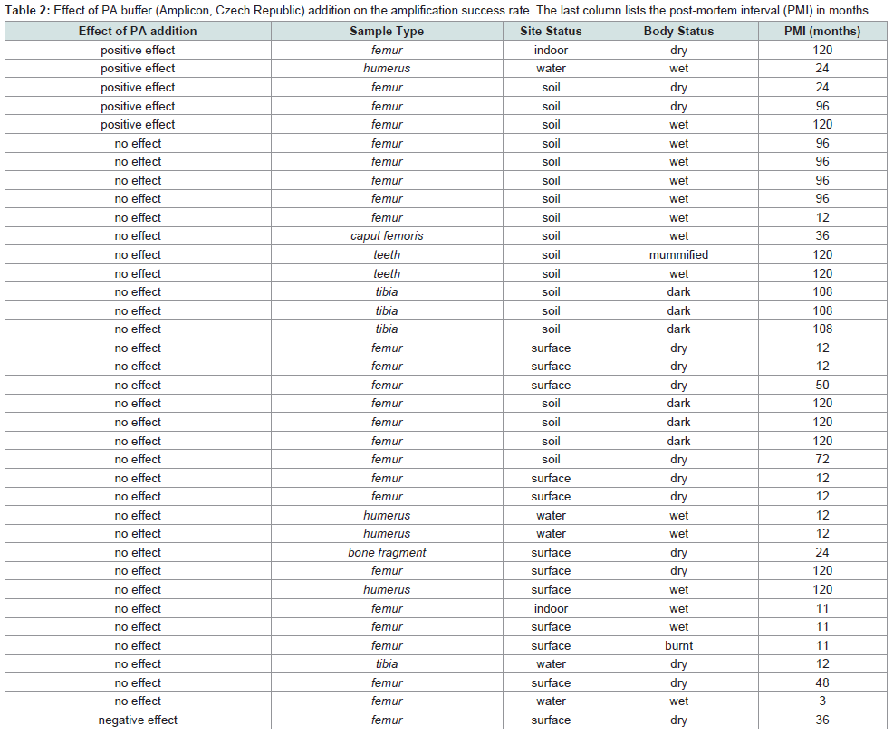
Table 2: Effect of PA buffer (Amplicon, Czech Republic) addition on the amplification success rate. The last column lists the post-mortem interval (PMI) in months.
The suitability of different forensic kits for human bone samples was tested to determine which kit could successfully analyze the DNA from human bone samples. The original IC-MP protocol (Davoren et al.) suggested the use of the PowerPlex16 kit.
The PowerPlex® 16 System (Promega Corporation) was used with a total of 166 samples, and its success rates are shown in Table 3. It has been shown that the PowerPlex 16 human identification kit (Promega Corporation) also amplifies non-human DNA of microbial origin. The presence of microbial DNA peaks in resulting DNA profiles was detected in 2 cases out of the 219 bodies analyzed. Example of microbial peak morphology is shown in Figure 11.
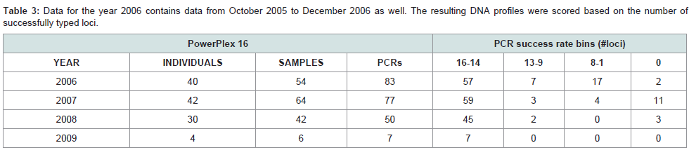
Table 3: Data for the year 2006 contains data from October 2005 to December 2006 as well. The resulting DNA profiles were scored based on the number of successfully typed loci.
Discussion
Aged bone samples are often considered to be among the most difficult forensic samples for DNA identification [1,2]. Therefore, it is important that DNA is extracted using a robust method that guarantees high yields of genetic material that is compatible with downstream processes, such as qRT-PCR and multiplex PCR.The identification of victims in mass graves or in mass fatalities such as the WTC event has been thoroughly covered in the scientific literature [2-6]. However, there are limited or no resources describing daily experiences with difficult criminal cases involving skeletal material. The data presented here comprise the period from October 2005 to December 2011, during which time the specialized police laboratory for DNA identification of bone samples processed a total of 323 samples from 219 bodies.
The number of tooth samples submitted increased each year, resulting in a significant reduction in the number of other sample types. This was a result of feedback provided to law enforcement agencies that submitted these samples. The quality of the samples submitted to the DNA laboratory during the years 2006-2008 significantly improved, mainly due to continued training and the availability of guidelines to law enforcement agencies. Close cooperation and communication with the Institute of the Legal Medicine also positively influenced the preparation of samples for submission, particularly the selection of the maceration technique [7,8] and sample storage prior to and during transport.
DNA extracted from decomposed human remains frequently contains fragmented human DNA as well as microbial DNA. Human DNA-specific extraction techniques are not available, particularly for very low quantities of DNA; thus, the presence of microbial DNA in extracts is unavoidable. An example of the negative effects of coextracted microbial DNA was demonstrated with Quantiblot DNA quantification [9]. Some widely used human forensic multiplexes have the ability to amplify various microbial DNAs, thus generating non-specific PCR products. By testing thousands of bone samples, the International Commission on Missing Persons (ICMP) has become aware of several noticeable patterns that are associated with various bacterial strains. These recognizable patterns are a clear indicator of the presence of bacterial peaks [10]. Additionally, the resulting GeneScan® profiles of suspected bacterial peaks exhibit an absence of artificial repeat slippage, sometimes referred to “n-4 bands,” “stutter” or “shadow bands.” Even if repeat sequences had been described in bacteria, yeast and fungi DNA, the detection of a “microbial peak” with a loss of a repeat unit has not yet been described.
The addition of PA buffer (Amplicon) to the extraction protocolwas tested since it would increase the binding efficiency of small DNA fragments to the silica matrix during extraction with guanidine salts. We observed that with PA buffer (Amplicon), 84% of the samples exhibited the same amplification success rate as without. The addition of PA buffer (Amplicon) had a positive effect for 14% of the samples, and only 1 sample (2%) exhibited a negative effect. We can conclude that the addition of PA buffer (Amplicon) has a slightly positive effect on the success rate, but it is not possible to decide in which case scenarios (PMI, site and body status) it should be recommended, as the marginally positive effects could be random.
Longer PMIs correspond with decreased amplification success rates. All samples from cases with PMIs shorter than 1 year gave positive results. This is clear evidence that the length of the postmortem interval influences the DNA identification success rate.
The PowerPlex® 16 System (Promega Corporation) and the AmpFlSTR® Identifiler™ (Applied Biosystems) system were directly compared for 14 samples. The results slightly favored the use of Identifiler rather than PowerPlex 16, but this conclusion was compromised by the limited number of samples for which a direct comparison was performed (see Table 4).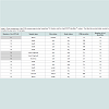
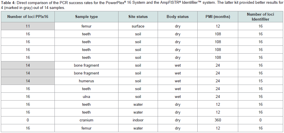
Table 4: Direct comparison of the PCR success rates for the PowerPlex® 16 System and the AmpFlSTR® Identifiler™ system. The latter kit provided better results for 4 (marked in gray) out of 14 samples.
Conclusion
The results presented above provide strong evidence that our laboratory set-up and protocols for DNA extraction from compact bones and teeth are optimal for bone samples submitted for forensic DNA analysis. These comprehensive guidelines for the set-up and operation of specialized bone identification DNA laboratories should serve as references for all planned or operational specialized laboratories for DNA identification from bone samples. It is important to note that the method described herein requires relatively large amount of material and thus the modification of the protocol consisting of the total demineralization step prior to the extraction enables to get DNA results from smaller and more difficult samples [11,12].Acknowledgements
We acknowledge the assistance of the Institute of Legal Medicine in Martin, Slovak Republic during the preparation of guidelines for bone sample submission as well as for their helpful comments during the preparation of this manuscript. We also acknowledge the Special Police Unit of the Slovak Republic for their tremendous help in securing funds for the start-up of this project. This work was supported by the Institute of Forensic Science of Slovak Police Forces.References
- Huffine E, Crews J, Kennedy B, Bomberger K, Zinbo A (2001) Mass identification of persons missing from the break-up of the former Yugoslavia: structure, function, and role of the International Commission on Missing Persons. Croat Med J 42: 271-275.
- Budimlija ZM, Prinz MK, Zelson-Mundorff A, Wiersema J, Bartelink E, et al. (2003) World Trade Center Human Identification Project: Experiences with Individual Body Identification Case. Croat Med J 44: 259-263.
- Davoren J, Vanek D, Konjhodzic R, Crews J, Huffine E, et al. (2007) Highly effective DNA extraction method for nuclear short tandem repeat testing of skeletal remains from mass graves. Croat Med J 48: 478-485.
- Miloš A, Selmanović A, Smajlović L, Huel R.L.M, Katzmarzyk Ch, et al. (2007) Success Rates of Nuclear Short Tandem Repeat Typing from Different Skeletal Elements. Croat Med J 48: 486-493.
- Palo JU, Hedman M, Söderholm N, Sajantila A (2007) Repatriation and Identification of Finnish World War II Soldiers. Croat Med J 48: 528-535.
- Marjanović D, Durmić-Pašić A, Bakal N, Haverić S, Kalamujić B, et al. (2007) DNA Identification of Skeletal Remains from World War II Mass Graves Uncovered in Slovenia. Croat Med J 48: 513-519.
- Steadman DW, Di Antonio LL, Wilson JJ, Sheridan KE, Tammariello SP (2006) The effects of chemical and heat maceration techniques on the recovery of nuclear and mitochondrial DNA from bone. J Forensic Sci 51: 11-17.
- Lee EJ, Luedtke JG, Allison JL, Arber CE, Merriwether DA, et al. (2010) The effects of different maceration techniques on nuclear DNA amplification using human bone. J Forensic Sci 55: 1032-1038.
- Alonso A, Anđelinović Š, Martín P, Sutlović D, Erceg I, et al. (2001) DNA Typing from Skeletal Remains: Evaluation of Multiplex and Megaplex STR Systems on DNA Isolated from Bone and Teeth Samples. Croat Med J 42: 260-266.
- Vanek D, Davoren J, Huffine E, Crews J, Konjhodzic R (2003) Products of Microbial DNA Amplification: Risks of False Results during DNA Typing of Decomposed Bodies and Skeletal Remains. Proceedings of the American Academy of Forensic Sciences 9.
- Amory S, Huel R, Bilić A, Loreille O, Parsons TJ (2012) Automatable full demineralization DNA extraction procedure from degraded skeletal remains. Forensic Sci Int Genet 6: 398-406.
- Vanek D, Saskova L, Koch H (2009) Kinship and Y-chromosome analysis of the 7th century human remains: novel DNA extraction and typing procedure for ancient material. Croat Med J 50: 286-295.