
Journal of Emergency Medicine & Critical Care
Download PDF
Case Report
*Address for Correspondence: Fadi T. Kasyouhanan, Emergency Medicine Chief Resident PGY- 3, Beaumont Health System, Ohio State University College of Medicine, University of Michigan-Ann Arbor, USA, E-mail: Fadi. Kasyouhanan@gmail.com
Citation: Singh S, Bahl A, Kasyouhanan FT. Rapid Resolution of Hydronephrosis after Spontaneous Passage of Kidney Stone. J Emerg Med Critical Care. 2015;1(2): 2.
Copyright © 2015 Singh S, et al. This is an open access article distributed under the Creative Commons Attribution License, which permits unrestricted use, distribution, and reproduction in any medium, provided the original work is properly cited.
Journal of Emergency Medicine & Critical Care| Volume: 1, Issue: 1
Submission: 13 August, 2015 | ISSN: 2469-4045 |Accepted: 04 September, 2015 | Published: 08 September, 2015
Reviewed & Approved by: Dr. Chad M. Cannon, Research Director, Department of Emergency Medicine, University of Kansas Hospital, Kansas, Missouri, USA
Rapid Resolution of Hydronephrosis after Spontaneous Passage of Kidney Stone
Sukhvir Singh, Amit Bahl and Fadi T. Kasyouhanan*
- Emergency Medicine Chief Resident PGY-3, Beaumont Health System, Ohio State University College of Medicine, University of Michigan-Ann Arbor, USA
*Address for Correspondence: Fadi T. Kasyouhanan, Emergency Medicine Chief Resident PGY- 3, Beaumont Health System, Ohio State University College of Medicine, University of Michigan-Ann Arbor, USA, E-mail: Fadi. Kasyouhanan@gmail.com
Citation: Singh S, Bahl A, Kasyouhanan FT. Rapid Resolution of Hydronephrosis after Spontaneous Passage of Kidney Stone. J Emerg Med Critical Care. 2015;1(2): 2.
Copyright © 2015 Singh S, et al. This is an open access article distributed under the Creative Commons Attribution License, which permits unrestricted use, distribution, and reproduction in any medium, provided the original work is properly cited.
Journal of Emergency Medicine & Critical Care| Volume: 1, Issue: 1
Submission: 13 August, 2015 | ISSN: 2469-4045 |Accepted: 04 September, 2015 | Published: 08 September, 2015
Reviewed & Approved by: Dr. Chad M. Cannon, Research Director, Department of Emergency Medicine, University of Kansas Hospital, Kansas, Missouri, USA
Abstract
Obstructing kidney stones can present with sudden onset of pain with varying degree of hydronephrosis. The timing of resolution of hydronephrosis after relief of obstruction has not been clearly demonstrated but can take days or even weeks for complete resolution. Long-standing hydronephrosis is associated with obstructive nephropathy and renal failure. We present a case of a patient with renal colic due to presumed obstructing stone with immediate complete resolution of hydronephrosis after presumed passage of a stone.Introduction
Patients with renal colic due to obstructing kidney stones often present with hydronephrosis on renal ultrasound or computed tomography (CT scan). Pain typically improves with resolution of obstruction by passage of stone. The timing of complete resolution of hydronephrosis after relief of obstruction is poorly understood. Resolution of hydronephrosis decreases a patient’s risk for obstructive nephropathy, renal failure, infection, and sepsis. We present a case showing moderate to severe hydronephrosis on ultrasonography in a patient presenting with acute right side pain. A CT scan was also completed within one hour of the ultrasound and revealed complete resolution of the hydronephrosis.Case Report
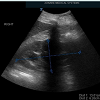
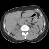
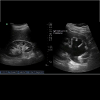
The patient is a 23-year-old female with a sudden onset of colicky right side abdominal/flank pain. The pain is associated with nausea, urinary urgency and frequency. There is no history of fever, chills, vomiting, dysuria, or hematuria. She has a history of “small kidney stones”. She has no history of vaginal discharge, vaginal bleeding, ovarian cysts, and no past surgeries. All of her vitals are within normal limits. She had mild tenderness in the right lower quadrant and mild tenderness over the right costovertebral angle. Urinalysis shows 1+ blood, 7 RBCs, 2 WBCs, negative for nitrites, leukocyte esterase, and bacteria. Pertinent labs include WBC of 7.6, creatinine of 0.63, and negative serum HCG. A point-of-care renal ultrasound scan is performed by the treating physician at 12:23 PM while the patient is symptomatic and reveals moderate to severe right sided hydronephrosis (Figure 1). A CT scan is completed at 1:18 PM and shows subtle edematous changes in the right kidney and no hydronephrosis or obstructing radiopaque stone (Figure 2). She reports that her pain has resolved prior to CT scan when she likely passed the presumed kidney stone (Figure 3). She is discharged home from the emergency department symptom free.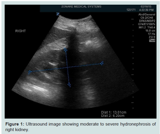
Figure 1: Ultrasound image showing moderate to severe hydronephrosis of right kidney.
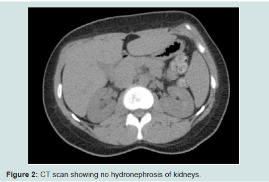
Figure 2:CT scan showing no hydronephrosis of kidneys.
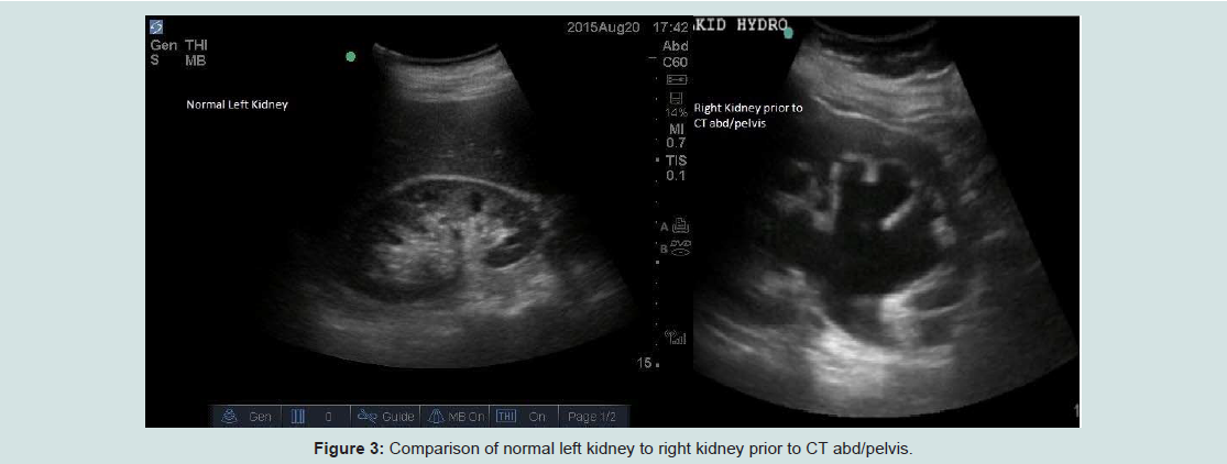
Figure 3: Comparison of normal left kidney to right kidney prior to CT abd/pelvis.